Forward and side scatter profile of human lymphocytes after peripheral blood centrifugation with Lymphopure™ (top dot plot), an equivalent product (middle plot), or no separation (bottom plot, lysed blood).
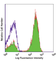
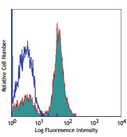
Histogram overlay shows CD3 or CD15 profile on total cells from lysed peripheral human blood before (dotted histogram) and after Lymphopure™ (purple tinted histogram) or equivalent product (orange open histogram) centrifugation. CD3 is a molecule expressed by T cells, which are recovered with the use of the isolation media. CD15 is a molecule highly expressed by granulocytes, which are absent after isolation with Lymphopure™.
Diagram showing whole blood cell separation using Lymphopure™. Blood is diluted with saline buffer and carefully layered over the medium in a conical tube, avoiding disturbing the interphase. Following centrifugation, at the appropriate speed with no braking, well-defined phases can be observed: 1-plasma, 2-interphase with mononuclear cells, 3-centrifugation medium, and 4-erythrocytes and granulocytes. Cells of interest can be aspirated from the interphase.
Cat #
Size
Price
Quantity
Check Availability
Save
Input string was not in a correct format.
426201
250 mL
$89
Input string was not in a correct format.
426202
500 mL
$153
Description
Lymphopure™ is a ready-to-use filtered and endotoxin tested solution for the isolation of mononuclear cells (MNCs) from a cell suspension. The solution is formulated at a density that allows separation of MNCs from other cells in blood such as granulocytes and erythrocytes. Granulocytes and erythrocytes will sediment after centrifugation.
Collect blood into a tube containing anti-coagulant (EDTA, heparin)
Dilute the blood with an equal volume of phosphate-buffered saline or similar suitable medium.
Mix Lymphopure™ by inverting the bottle several times.
Aliquot half the amount of Lymphopure™ into a tube. For every ml of diluted blood, add 0.5 ml of Lymphopure™.
Carefully layer the diluted blood on top of Lymphopure™. Avoid mixing blood with Lymphopure™.
Centrifuge at 800 x g for 20 minutes at room temperature (15 - 25°C) without braking. If the blood has been stored for more than 2 hours, centrifuge for 30 minutes.
After centrifugation, mononuclear cells (MNCs) form a defined cell layer at the plasma: Lymphopure™ interface.
Collect the MNC layer at the interface without disturbing the other fractions.
Wash MNCs with medium.
Application References
(PubMed link indicates BioLegend citation)
Böyum A. 1968. Scand. J. Clin. Lab. Invest. Suppl. 97:9-29.
Böyum A. 1968. Scand. J. Clin. Lab. Invest. Suppl. 97:77-89.
Böyum A. 1964. Nature. 204:793-4.
Thorsby E, Bratile A. 1970. Histocompatibility Testing 1970,ed. P.I. Terasaki. 655-6.
 Login/Register


Follow Us