- Clone
- W20045D (See other available formats)
- Regulatory Status
- RUO
- Other Names
- Excision Repair Cross-Complementation Group 1, ERCC excision repair 1, endonuclease non-catalytic subunit, RAD10
- Isotype
- Rat IgG2b, κ
- Ave. Rating
- Submit a Review
- Product Citations
- publications

-

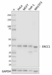
Whole cell extracts (15 µg total protein) from HeLa, MCF7, THP-1, and NIH/3T3 cells were resolved by 4-12% Bis-Tris gel electrophoresis, transferred to a PVDF membrane, and probed with 0.25 μg/mL (1:2000 dilution) of purified anti-ERCC1 antibody (clone W20045D) overnight at 4°C. Proteins were visualized by chemiluminescence detection using HRP goat anti-rat IgG antibody (Cat. No. 405405) at a 1:3000 dilution. Direct-Blot™ HRP anti-GAPDH antibody (Cat. No. 607904) was used as a loading control at a 1:50000 dilution (lower). Western-Ready™ ECL Substrate Premium Kit (Cat. No. 426319) was used as a detection agent. Lane M: Molecular weight marker. -

ICC staining using purified anti-ERCC1 antibody (clone W20045D) on HeLa cells. The cells were fixed and permeabilized with 100% methanol, and blocked with 5% FBS for 1 hour. The cells were then stained with either no primary antibody (panel A) or 0.625 µg/mL of the primary antibody (panel B), followed by incubation with 2.5 µg/mL of Alexa Fluor® 594 anti-goat anti-rat IgG antibody (Cat. No. 405422) for 1 hour at room temperature. Nuclei were counterstained with DAPI, and the images were captured with a 60X objective. -

IHC staining of anti-ERCC1 antibody (clone W20045D) on formalin-fixed paraffin-embedded human prostate cancer (A, positive control) and normal prostate (B, negative control) tissues. Following antigen retrieval using Sodium Citrate H.I.E.R. (Cat. No. 928502), the tissues were incubated with 5 µg/mL of anti-ERCC1 antibody overnight at 4°C. BioLegend’s Ultra Streptavidin HRP Kit (Multi-Species, DAB, Cat. No. 929501) was used for detection followed by hematoxylin counterstaining, according to the protocol provided. The images were captured with a 40X objective. Scale bar: 50 μm.
| Cat # | Size | Price | Quantity Check Availability | Save | ||
|---|---|---|---|---|---|---|
| 947501 | 25 µg | $118 | ||||
| 947502 | 100 µg | $293 | ||||
ERCC1, also referred to as Excision Repair Cross-Complementation Group 1, is a component of the nucleotide excision repair pathway. ERCC1 forms a heterodimer with XPF endonuclease to catalyze the 5’ incision in the process of excising a DNA lesion. ERCC1 also works with SLX4 in homology direct repair of double strand breaks and in the repair of crosslinks. Mutations in ERCC1 play a role in carcinogenesis due to genome instability, and elevated expression of ERCC1 is predictive of resistance to platinum-based chemotherapies in multiple types of cancers.
Product DetailsProduct Details
- Verified Reactivity
- Human, Mouse
- Antibody Type
- Monoclonal
- Host Species
- Rat
- Immunogen
- Partial recombinant human ERCC1 protein
- Formulation
- Phosphate-buffered solution, pH 7.2, containing 0.09% sodium azide
- Preparation
- The antibody was purified by affinity chromatography.
- Concentration
- 0.5 mg/mL
- Storage & Handling
- The antibody solution should be stored undiluted between 2°C and 8°C.
- Application
-
WB - Quality tested
IHC-P, ICC - Verified - Recommended Usage
-
Each lot of this antibody is quality control tested by western blotting. For western blotting, the suggested use of this reagent is 0.125 - 1.0 µg/mL. For immunohistochemistry on formalin-fixed paraffin-embedded tissue sections, a concentration range of 5.0 - 10.0 µg/mL is suggested. For immunocytochemistry, a concentration range of 0.625 - 5.0 μg/mL is recommended. It is recommended that the reagent be titrated for optimal performance for each application.
- Application Notes
-
Clone W20045D detects multiple ERCC1 isoforms by western blot.
Clone W20045D was tested for ICC using HeLa cells fixed with methanol or 4% paraformaldehyde and permeabilized with either methanol or Triton X-100. All three methods were compatible with ERCC1 staining. - RRID
-
AB_2894534 (BioLegend Cat. No. 947501)
AB_2894534 (BioLegend Cat. No. 947502)
Antigen Details
- Structure
- ERCC1 is a 297 amino acid protein with an expected molecular weight of 33 kD. Four isoforms between 25 and 36 kD have been described.
- Distribution
-
Ubiquitously expressed/ Nucleus
- Function
- DNA damage repair
- Biology Area
- Cell Biology, DNA Repair/Replication
- Molecular Family
- Enzymes and Regulators
- Antigen References
-
- Friboulet, et al. 2013. Cell Cycle. 12:3298
- Kashiyama, et al. 2013. J Hum Genet. 92:807
- Wan J, et al. 2017. Canc Invest. 35:85
- Faridounnia, et al. 2018. Molecules. 23:3205
- Gene ID
- 2067 View all products for this Gene ID
- UniProt
- View information about ERCC1 on UniProt.org
Related Pages & Pathways
Pages
Related FAQs
Other Formats
View All ERCC1 Reagents Request Custom Conjugation| Description | Clone | Applications |
|---|---|---|
| Purified anti-ERCC1 | W20045D | WB,IHC-P,ICC |
Compare Data Across All Formats
This data display is provided for general comparisons between formats.
Your actual data may vary due to variations in samples, target cells, instruments and their settings, staining conditions, and other factors.
If you need assistance with selecting the best format contact our expert technical support team.
 Login/Register


Follow Us