- Clone
- W15090A (See other available formats)
- Regulatory Status
- RUO
- Other Names
- Histone deacetylase-like 1 (HDACL1), MRXS6, Wilson-Turner X-linked mental retardation syndrome (WTS), RPD3
- Isotype
- Mouse IgG1, κ
- Ave. Rating
- Submit a Review
- Product Citations
- publications

-

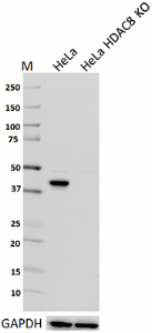
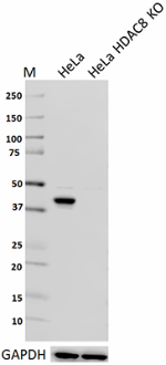
Total lysates (10µg protein) from HeLa and HeLa HDAC8 CRISPR/Cas9 knockout (KO) cells were resolved by electrophoresis (4-20% Tris-glycine gel), transferred to nitrocellulose, and probed with 1:500 (1µg/ml) purified anti-HDAC8 antibody , clone W15090A (upper) or 1:3000 diluted anti-GAPDH (poly6314, cat#631401) antibody (lower). Proteins were visualized using chemiluminescence detection by incubation with HRP goat anti-mouse-IgG secondary antibody (cat#405306) for the anti-HDAC8 antibody, and a donkey anti-rabbit IgG Antibody conjugated to HRP (Cat# 406401) for GAPDH. Lane M is the Molecular weight ladder. -

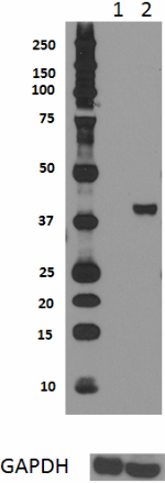
Total lysates (15 µg protein) mouse brain (lane 1) and HeLa cells (lane 2) were resolved by electrophoresis (4-12% Bis-Tris gel), transferred to nitrocellulose, and probed with 1:1000 diluted (0.5 µg/mL) Purified anti-HDAC8 Antibody, clone W15090A (upper) or 1:3000 diluted Purified anti-GAPDH Antibody, clone poly6314 (lower). Proteins were visualized by chemiluminescence detection using a 1:3000 diluted goat anti-mouse-IgG secondary antibody conjugated to HRP for the anti-HDAC8 Antibody, and a donkey anti-rabbit IgG Antibody conjugated to HRP for anti-GAPDH Antibody. -

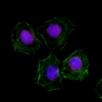
HeLa cells were fixed with 2% paraformaldehyde (PFA) for 15 minutes, permeabilized with 0.5% Triton X-100 for three minutes, and blocked with 1% BSA for 60 minutes. Then the cells were intracellularly stained with 2 µg/ml purified anti-human HDAC8 (clone W15090A) antibody for one hour, followed by staining with Alexa Fluor® 594 anti-mouse IgG antibody (red), Alexa Fluor® 488 Phalloidin (green), and DAPI (blue) for one hour at room temperature. The image was captured with a 60X objective. -

Chromatin Immunoprecipitation (ChIP) was performed using commercial Protein-G coated 96 well high-throughput ChIP assay kit by loading 3µg of cross-linked chromatin samples from Jurkat cells with either A) 1:50 dilution of Go-ChIP-Grade™ Purified anti-HDAC8 Antibody (Clone W15090A), B) equal amount of Purified Mouse IgG1, κ Isotype Control Antibody, or C) competitor’s ChIP-grade Purified anti-HDAC8 Antibody and D) equal amount of matched Isotype Control Antibody as recommended by the manufacturer. The enriched DNA was purified and quantified by real-time qPCR using primers targeting human EGR-1 gene region. The amount of immunoprecipitated DNA in each sample is represented as signal relative to the 5% of total amount of input chromatin.
| Cat # | Size | Price | Quantity Check Availability | Save | ||
|---|---|---|---|---|---|---|
| 685502 | 100 µg | $311 | ||||
Histone deacetylases (HDACs) are a family of enzymes responsible for the removal of acetyl groups from lysine residues on the core histones and other non-histone proteins. HDAC8 is a type I histone deacetylase and was initially found to deacetylate a variety of acetylated histone variants in vitro. HDAC8 is a kinectically and structurally well-characterized deacetylase that was reported to catalyze several nuclear proteins, which include ERR-α, hEST1B, and SMC3. Patients with HDAC8 mutations show Cornelia de Lange Syndrome (CdLS) and Wilson-turner Syndrome (WTS) phenotypes. Elevated expression of HDAC8 has been observed in various tumor types, such as acute lymphoblastic leukemia (AML), neuroblastomas, and glimoas. HDAC8 is implicated as a therapeutic target in cancer and infectious diseases.
Product DetailsProduct Details
- Verified Reactivity
- Human
- Antibody Type
- Monoclonal
- Host Species
- Mouse
- Immunogen
- Full length human HDAC8 recombinant protein expressed in E. coli.
- Formulation
- Phosphate-buffered solution, pH 7.2, containing 0.09% sodium azide.
- Preparation
- The antibody was purified by affinity chromatography.
- Concentration
- 0.5 mg/ml
- Storage & Handling
- The antibody solution should be stored undiluted between 2°C and 8°C.
- Application
-
WB - Quality tested
KO/KD-WB, ICC, ChIP - Verified - Recommended Usage
-
Each lot of this antibody is quality control tested by Western blotting. For Western blotting, the suggested use of this reagent is 0.5 - 2.0 µg per ml. For immunocytochemistry, a concentration range of 1.0 - 5.0 μg/ml is recommended. For ChIP applications, the suggested dilution is 1:50 by volume. It is recommended that the reagent be titrated for optimal performance for each application.
- Product Citations
-
- RRID
-
AB_2572204 (BioLegend Cat. No. 685502)
Antigen Details
- Structure
- 377 amino acids with a predicted molecular weight of 41.8 kD. It belongs to the type I Histone deacetylase subfamily.
- Distribution
-
Nucleus and cytoplasm.
- Function
- HDAC8 is a deacetylase and regulates gene expression through catalyzing the removal of acetyl groups on lysine residues in the core histones and transcriptional factors.
- Interaction
- Interacts with PEPB2-MYH11, CBFA2T3, SMG5, and alpha-SMA.
- Biology Area
- Cell Biology, Chromatin Remodeling/Epigenetics, Neuroscience, Transcription Factors
- Antigen References
-
1. Qi J, et al. 2015. Cell Stem Cell. 17:597.
2. Tian Y, et al. 2015. Cancer Res. 75:4803.
3. Feng L, et al. 2015. J. Hum. Genet. 60:165.
4. Ha SD, et al. 2014. J. Immunol. 193:1333.
5. Higuchi T, et al. 2013. Blood 121:3640.
6. Deardorff MA, et al. 2012. Nature 489:313. - Gene ID
- 55869 View all products for this Gene ID
- UniProt
- View information about HDAC8 on UniProt.org
Related Pages & Pathways
Pages
Related FAQs
Other Formats
View All HDAC8 Reagents Request Custom Conjugation| Description | Clone | Applications |
|---|---|---|
| Purified anti-HDAC8 | W15090A | WB,KO/KD-WB,ICC,ChIP |
Compare Data Across All Formats
This data display is provided for general comparisons between formats.
Your actual data may vary due to variations in samples, target cells, instruments and their settings, staining conditions, and other factors.
If you need assistance with selecting the best format contact our expert technical support team.
 Login/Register


Follow Us