 Login/Register


- Clone
- W17240D (See other available formats)
- Regulatory Status
- RUO
- Other Names
- Protein Tyrosine Phosphatase, Non-Receptor Type 6, PTPN6, Hematopoietic Cell Protein-Tyrosine Phosphatase, Protein-Tyrosine Phosphatase SHP-1, Protein-Tyrosine Phosphatase 1C
- Isotype
- Rat IgG2b, κ
- Ave. Rating
- Submit a Review
- Product Citations
- publications

-

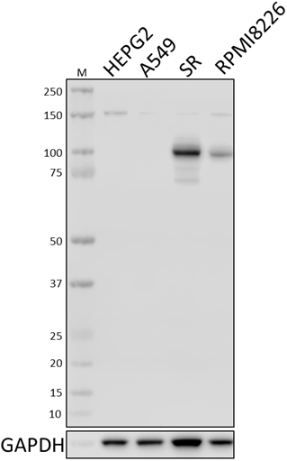
Whole cell extracts (15 µg protein) from SH-SY5Y and U87-MG (negative control), MOLT-4, Jurkat, THP-1, and EL4 cells were resolved by 4-12% Bis-Tris gel electrophoresis, transferred to a PVDF membrane, and probed with 0.1 µg/mL (1:5000 dilution) of Purified anti-SHP-1 Antibody, clone W17240D, overnight at 4°C. Proteins were visualized by chemiluminescence detection using HRP goat anti-rat IgG Antibody (Cat. No. 405405) at a 1:3000 dilution. Direct-Blot™ HRP anti-GAPDH Antibody (Cat. No. 607904) was used as a loading control at a 1:25000 dilution (lower). Lane M: Molecular Weight marker. Predicted expression data was obtained from Human Protein Atlas. -

Jurkat cells were fixed with 4% paraformaldehyde for 10 minutes, permeabilized with ice-cold methanol for 10 minutes, and blocked with 5% FBS for 60 minutes. Cells were then intracellularly stained with a 1:100 dilution (5.0 µg/mL) of either Purified Rat IgG2b, κ Isotype Control Antibody (Cat. No. 400602, panel A) or Purified anti-SHP-1 Antibody, clone W17240D (panel B) overnight at 4°C, followed by incubation with Alexa Fluor® 594 goat anti-rat IgG (Cat. No. 405422) at 2.0 µg/mL. Nuclei were counter-stained with DAPI, and the image was captured with a 60X objective. -

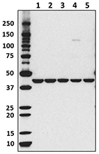
Whole cell extracts (250 µg total protein) prepared from Jurkat cells were immunoprecipitated overnight with 2.5 µg of Purified rat IgG2b, κ Isotype Control Antibody (Cat. No. 400602) or Purified anti-SHP-1 Antibody, clone W17240D. The resulting IP fractions and whole cell extract input (5%) were resolved by 4-12% Bis-Tris gel electrophoresis, transferred to a PVDF membrane, and probed with a 1.0 µg/mL (1:500 dilution) of W17240D. Lane M: Molecular Weight marker. -

K562 cells (negative control, open histogram) or Jurkat cells (positive control, filled histogram) were fixed and permeabilized using Cyto-Fast™ Fix/Perm buffer set (Cat. No. 426803) and intracellularly stained with purified anti-SHP-1 (clone W17240D), or purified Rat IgG2b, κ isotype control (open histogram, dashed line) (representative histogram for both cell lines) (Cat. No. 400601), followed by Alexa Fluor™ 647 goat anti-Rat IgG (Cat. No. 405416)
| Cat # | Size | Price | Quantity Check Availability | Save | ||
|---|---|---|---|---|---|---|
| 620301 | 25 µg | $101 | ||||
| 620302 | 100 µg | $253 | ||||
SHP-1, also referred to as PTPN6 is an inhibitory protein phosphatase widely expressed in hematopoietic cell lineages that regulates cell cycle, proliferation, and inflammatory pathways. SHP-1 dephosphorylates activating signaling molecules in receptor tyrosine kinase pathways; in T cells it functions as a negative regulator of T cell activation and proliferation. SHP-1 is constitutively bound to the ITIM-containing adaptor molecules such as the inhibitory receptor LAIR-1; activation of SHP-1 results in either direct dephosphorylation of the T cell receptor or downstream effector molecules such as Zap70 and Lck. Reduced expression of SHP1 is found in leukemias, lymphomas, and colorectal cancers due to promoter hypermethylation of the PTPN6 gene.
Product DetailsProduct Details
- Verified Reactivity
- Human
- Antibody Type
- Monoclonal
- Host Species
- Rat
- Immunogen
- Partial recombinant protein corresponding to amino acid residues 1-213 of human SHP-1
- Formulation
- Phosphate-buffered solution, pH 7.2, containing 0.09% sodium azide.
- Preparation
- The antibody was purified by affinity chromatography.
- Concentration
- 0.5 mg/mL
- Storage & Handling
- The antibody solution should be stored undiluted between 2°C and 8°C.
- Application
-
WB - Quality tested
ICC, IP, ICFC - Verified - Recommended Usage
-
Each lot of this antibody is quality control tested by Western blotting. For Western blotting, the suggested use of this reagent is 0.25 - 1.0 µg per mL. For immunocytochemistry, a concentration range of 1.0 - 5.0 μg/mL is recommended. For immunoprecipitation, the suggested use of this reagent is 2.5 µg per mL. For intracellular flow cytometric staining, the suggested use of this reagent is ≤ 0.25 µg per million cells in 100 µL volume. It is recommended that the reagent be titrated for optimal performance for each application.
- Application Notes
-
W17240D was tested for ICC using PFA-fixed Jurkat cells permeabilized with either Triton X-100 or methanol. While both permeabilization methods facilitated SHP-1 staining, methanol resulted in considerably stronger staining.
- Product Citations
-
- RRID
-
AB_2810679 (BioLegend Cat. No. 620301)
AB_2810679 (BioLegend Cat. No. 620302)
Antigen Details
- Structure
- SHP-1 is a 595 amino acid protein with a predicted molecular weight of 68 kD.
- Distribution
-
Cytoplasm and nucleus/hematopoietic cells
- Cell Type
- T cells, Thymocytes
- Biology Area
- Cell Biology, Cell Proliferation and Viability, Immuno-Oncology, Immunology
- Molecular Family
- Protein Kinases/Phosphatase
- Antigen References
-
- Paulson RF, et al. 1996. Nat Genet. 13:309.
- Khoury JD, et al. 2004. Blood. 104:1580.
- Paster W, et al. 2015. EMBO J. 34:393.
- Gene ID
- 5777 View all products for this Gene ID
- UniProt
- View information about SHP-1 on UniProt.org

Related FAQs
Other Formats
View All SHP-1 Reagents Request Custom Conjugation| Description | Clone | Applications |
|---|---|---|
| Purified anti-SHP-1 | W17240D | WB,ICC,IP,ICFC |
Customers Also Purchased


Compare Data Across All Formats
This data display is provided for general comparisons between formats.
Your actual data may vary due to variations in samples, target cells, instruments and their settings, staining conditions, and other factors.
If you need assistance with selecting the best format contact our expert technical support team.


Follow Us