- Clone
- S11 (See other available formats)
- Regulatory Status
- RUO
- Other Names
- Ly-48
- Isotype
- Rat IgG2b, κ
-

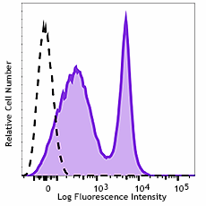
C57BL/6 mouse splenocytes were stained with anti-mouse CD43 (clone S11) Alexa Fluor® 700 (filled histogram) or rat IgG2b Alexa Fluor® 700 isotype control (open histogram).
| Cat # | Size | Price | Quantity Check Availability | ||
|---|---|---|---|---|---|
| 143213 | 25 µg | $118.00 | |||
| 143214 | 100 µg | $265.00 | |||
CD43, also known as Leukosialin and Ly48, is a 125 kD sialoprotein (glycosylated protein) expressed from 1.2 kBase mRNA in bone marrow-derived cells. This occurs early in development. Cells expressing CD43 include γ/δ T cells, macrophages, mature B cells, and dendritic cells. CD43 functions as an anti-adhesive surface molecule, promoted by antibody cross-linking, that releases the trailing edge of the cell during locomotion to allow movement of the cell body towards the lamellipodia. The intracellular distribution of CD43 is determined by binding to moesin, an intracellular membrane protein, which is in-turn bound in some manner to the actin cytoskeleton. Defects with CD43 function and expression retard cellular locomotion, resulting in a wide range of immune disorders. Wiscott-Alderich syndrome, and the varying degrees of its severity, is related to the dysregulation of CD43 expression.
Product Details
- Verified Reactivity
- Mouse
- Antibody Type
- Monoclonal
- Host Species
- Rat
- Immunogen
- Mouse plasmacytoma cells
- Formulation
- Phosphate-buffered solution, pH 7.2, containing 0.09% sodium azide.
- Preparation
- The antibody was purified by affinity chromatography and conjugated with Alexa Fluor® 700 under optimal conditions.
- Concentration
- 0.5 mg/ml
- Storage & Handling
- The antibody solution should be stored undiluted between 2°C and 8°C, and protected from prolonged exposure to light. Do not freeze.
- Application
-
FC - Quality tested
- Recommended Usage
-
Each lot of this antibody is quality control tested by immunofluorescent staining with flow cytometric analysis. For flow cytometric staining, the suggested use of this reagent is ≤ 0.5 µg per million cells in 100 µl volume. It is recommended that the reagent be titrated for optimal performance for each application.
* Alexa Fluor® 700 has a maximum emission of 719 nm when it is excited at 633 nm / 635 nm. Prior to using Alexa Fluor® 700 conjugate for flow cytometric analysis, please verify your flow cytometer's capability of exciting and detecting the fluorochrome.
Alexa Fluor® and Pacific Blue™ are trademarks of Life Technologies Corporation.
View full statement regarding label licenses - Excitation Laser
-
Red Laser (633 nm)
- Application Notes
-
Additional reported applications (for the relevant formats) include: Western blotting3. The S11 antibody reacts with pan-CD43.
-
Application References
(PubMed link indicates BioLegend citation) -
- Gaspari AA, et al. 1993. J. Invest. Dermatol. 100:247. (FC)
- Merzaban JS, et al. 2005. J. Immunol. 174:4051. (FC)
- Baecher-Allan CM, et al. 1993. Immunogenetics. 37:183. (WB)
- Product Citations
-
- RRID
-
AB_2800661 (BioLegend Cat. No. 143213)
AB_2800661 (BioLegend Cat. No. 143214)
Antigen Details
- Structure
- Single chain of type I transmembrane glycoprotein with abundant O-glycosylation and sialylation sites; 115 kD and 130 kD glycoforms with different glycosylation and sialylation.
- Distribution
-
Gamma/delta T cells, macrophages, mature B cells, and dendritic cells.
- Function
- Plays dual roles in cell-cell adhesion and anti-adhesion, costimulation of cell activation and survival, and induction of apoptosis of T cells and hematopoietic progenitors.
- Interaction
- CD43 functions as an anti-adhesive surface molecule, promoted by antibody cross-linking, that releases the trailing edge of the cell during locomotion to allow movement of the cell body towards the lamellipodia.
- Ligand/Receptor
-
Moesin, CD54, E-selectin, Siglec-1
. - Cell Type
- B cells, Dendritic cells, Macrophages, T cells
- Biology Area
- Cell Adhesion, Cell Biology, Cell Motility/Cytoskeleton/Structure, Immunology
- Molecular Family
- Adhesion Molecules, CD Molecules
- Antigen References
-
1. Van den Berg TK, et al. 2001. J. Immunol. 166:3637.
2. Moore T, et al. 1994. J. Immunol. 153:4978.
3. Onami TM, et al. 2002. J. Immunol. 168:6022.
4. Tong J, et al. 2004. J. Exp. Med. 199:1277.
5. Jones AT, et al. 1994. J. Immunol. 153:3426.
6. Matsumoto M, et al. 2005. J. Immunol. 175:8042. - Gene ID
- 20737 View all products for this Gene ID
- UniProt
- View information about CD43 on UniProt.org
Other Formats
View All CD43 Reagents Request Custom Conjugation| Description | Clone | Applications |
|---|---|---|
| APC anti-mouse CD43 | S11 | FC |
| PE/Cyanine7 anti-mouse CD43 | S11 | FC |
| Purified anti-mouse CD43 | S11 | FC,WB |
| FITC anti-mouse CD43 | S11 | FC |
| PE anti-mouse CD43 | S11 | FC |
| TotalSeq™-A0110 anti-mouse CD43 | S11 | PG |
| APC/Fire™ 750 anti-mouse CD43 | S11 | FC |
| Alexa Fluor® 700 anti-mouse CD43 | S11 | FC |
| PE/Dazzle™ 594 anti-mouse CD43 | S11 | FC |
| PerCP/Cyanine5.5 anti-mouse CD43 | S11 | FC |
| TotalSeq™-C0110 anti-mouse CD43 | S11 | PG |
| TotalSeq™-B0110 anti-mouse CD43 | S11 | PG |
Compare Data Across All Formats
This data display is provided for general comparisons between formats.
Your actual data may vary due to variations in samples, target cells, instruments and their settings, staining conditions, and other factors.
If you need assistance with selecting the best format contact our expert technical support team.
-
APC anti-mouse CD43
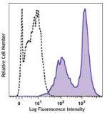
Mouse splenocytes were stained with CD43 (clone S11) APC (fi... -
PE/Cyanine7 anti-mouse CD43
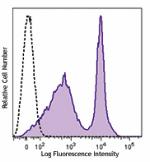
C57BL/6 mouse splenocytes were stained with either anti-mous... -
Purified anti-mouse CD43
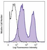
C57BL/6 mouse splenocytes were stained with either purified ... -
FITC anti-mouse CD43
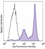
C57BL/6 mouse splenocytes were stained with either anti-mous... -
PE anti-mouse CD43
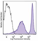
C57BL/6 mouse splenocytes were stained with either anti-mous... -
TotalSeq™-A0110 anti-mouse CD43
-
APC/Fire™ 750 anti-mouse CD43
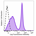
C57BL/6 mouse splenocytes were stained with anti-mouse CD43 ... -
Alexa Fluor® 700 anti-mouse CD43
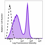
C57BL/6 mouse splenocytes were stained with anti-mouse CD43 ... -
PE/Dazzle™ 594 anti-mouse CD43
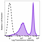
C57BL/6 mouse splenocytes were stained with anti-mouse CD43 ... -
PerCP/Cyanine5.5 anti-mouse CD43
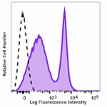
C57BL/6 mouse splenocytes were stained with anti-mouse CD43 ... -
TotalSeq™-C0110 anti-mouse CD43
-
TotalSeq™-B0110 anti-mouse CD43
