- Clone
- A53-B/A2 (See other available formats)
- Regulatory Status
- RUO
- Other Names
- Keratin 19, Keratin 19 type 1 cytoskeletal
- Isotype
- Mouse IgG2a, κ
-
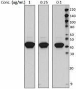
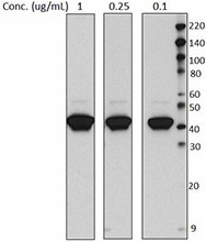
Total cell lysate (15 µg protein) from MCF-7 were resolved by 4-20% Tris-glycine gel electrophoresis, transferred to nitrocellulose, and probed with different concentrations of purified anti-Cytokeratin 19 Antibody (clone A53-B/A2). Proteins were visualized using a goat anti-mouse-IgG secondary antibody conjugated to HRP and chemiluminescence detection -

MCF-7 cells were stained with anti-Cytokeratin 19 (clone A53-B/A2), followed by Alexa Fluor® 546 secondary antibody and DAPI (nuclei). Images were aquired on a Nikon FC300 inverted microscope at 20X magnification. Data provided by Dr. John Nolan, La Jolla Bioengineering Institute. -

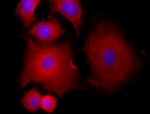
MCF-7 cells were stained with purified anti- Cytokeratin 19 (A53-B/A2) antibody, followed by staining with DyLight™ 594 conjugated goat anti-mouse IgG (red) antibody. Nuclei were stained with DAPI (blue).
| Cat # | Size | Price | Quantity Check Availability | ||
|---|---|---|---|---|---|
| 628502 | 100 µg | $218.00 | |||
Cytokeratin 19, also known as keratin 19, is a type I intermediate filament protein with a molecular weight of approximately 40-44 kD. Cytokeratin 19 is a heterotetramer composed of two type I and two type II keratin subunits. Unlike other cytokeratins, cytokeratin 19 lacks a C-terminal non-helical extension. This cytokeratin is widely expressed in the periderm (transient superficial layer enveloping developing epidermis), muscle, intestine, bile duct, esophagus, stomach, and thymus. Cytokeratin 19 can be upregulated by vitamin A and is thought to play a critical role in embryogenesis. Cytokeratin 19 intereacts with the pinin protein and has been shown to be modified by phosphorylation (Ser10, Ser35).
Product Details
- Verified Reactivity
- Human
- Antibody Type
- Monoclonal
- Host Species
- Mouse
- Immunogen
- Human mammary carcinoma cell line MCF-7
- Formulation
- This antibody is provided in phosphate-buffered solution, pH 7.2, containing 0.09% sodium azide at 0.5 mg/ml.
- Preparation
- The antibody was purified by affinity chromatography.
- Concentration
- 0.5 mg/ml
- Storage & Handling
- The antibody solution should be stored undiluted between 2°C and 8°C.
- Application
-
WB - Quality tested
ICC - Verified
IP, IHC-P, ELISA - Reported in the literature, not verified in house - Recommended Usage
-
Each lot of this antibody is quality control tested by Western blotting. Western blotting, suggested working dilution(s): Use 5 µg antibody per 5 ml antibody dilution buffer for each mini-gel. It is recommended that the reagent be titrated for optimal performance for each application.
- Application Notes
-
Additional reported applications (for the relevant formats) include: immunoprecipitation, immunohistochemistry of paraffin-embedded sections, immunocytochemistry, and ELISA.
-
Application References
(PubMed link indicates BioLegend citation) -
- Karsten U, et al. 1985. Eur. J. Cancer Clin. Oncol. 21:733.
- Nishikata T,et al. Anticancer Res. 33:2867. PubMed
- Product Citations
-
- RRID
-
AB_439773 (BioLegend Cat. No. 628502)
Antigen Details
- Structure
- Type I intermediate filament protein, contains three coiled-coil domains, lacks C-terminal non-helical extension found in other keratins, approximately 40-44 kD. Cytokeratin 19 is a heterotetramer composed of two type I and two type II keratin subunits.
- Distribution
-
Expressed in the periderm (transient superficial layer enveloping developing epidermis), also expressed in muscle, intestine, bile duct, esophagus, stomach, and thymus. Upregulated by vitamin A.
- Function
- Intermediate filament protein involved with the cytoskeleton. May play a critical role in embryogenesis
- Interaction
- Interacts with pinin
- Modification
- Phosphorylation (Ser10, Ser35)
- Biology Area
- Cell Biology, Cell Motility/Cytoskeleton/Structure, Neuroscience, Neuroscience Cell Markers
- Molecular Family
- Intermediate Filaments
- Antigen References
-
1. Bader BL, et al. 1986. EMBO J. 5:1865.
2. Eckert RL. 1988. Proc. Natl. Acad. Sci. 85:1114.
3. Stasiak PC and Lane EB. 1987 Nucleic Acids Res. 15:10058. - Gene ID
- 3880 View all products for this Gene ID
- UniProt
- View information about Cytokeratin 19 on UniProt.org
Other Formats
View All Cytokeratin Reagents Request Custom Conjugation| Description | Clone | Applications |
|---|---|---|
| Purified anti-Cytokeratin 19 | A53-B/A2 | WB,ICC,IP,IHC-P,ELISA |
| Alexa Fluor® 594 anti-Cytokeratin 19 | A53-B/A2 | ICC,IHC-P |
| Alexa Fluor® 647 anti-Cytokeratin 19 | A53-B/A2 | ICC |
| Alexa Fluor® 488 anti-Cytokeratin 19 | A53-B/A2 | ICC |
Compare Data Across All Formats
This data display is provided for general comparisons between formats.
Your actual data may vary due to variations in samples, target cells, instruments and their settings, staining conditions, and other factors.
If you need assistance with selecting the best format contact our expert technical support team.
-
Purified anti-Cytokeratin 19
Total cell lysate (15 µg protein) from MCF-7 were resolved ... 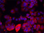
MCF-7 cells were stained with anti-Cytokeratin 19 (clone A5... 
MCF-7 cells were stained with purified anti- Cytokeratin 19 ... -
Alexa Fluor® 594 anti-Cytokeratin 19
MCF7 cells were fixed with 1% paraformaldehyde (PFA), permea... 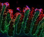
Human paraffin-embedded colon was prepared with standard dep... -
Alexa Fluor® 647 anti-Cytokeratin 19
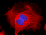
MCF-7 cells were fixed with 1% paraformaldehyde (PFA) for 10... -
Alexa Fluor® 488 anti-Cytokeratin 19
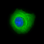
MCF-7 cells were fixed with 1% paraformaldehyde (PFA) for te...


