- Clone
- DA-7 (See other available formats)
- Regulatory Status
- RUO
- Other Names
- Keratin 18, Keratin type I cytoskeletal 18
- Isotype
- Mouse IgG1, κ
- Ave. Rating
- Submit a Review
- Product Citations
- publications
-

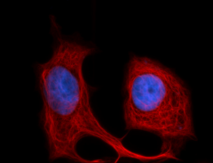
MCF-7 cells were fixed with 1% paraformaldehyde (PFA) for 10 minutes at 37°C, permeabilized with 0.5% Triton X-100 buffer for 10 minutes at room temperature, and blocked for 30 minutes at room temperature with 5% fetal bovine serum (FBS). Then the cells were stained with 5 µg/mL anti-Cytokeratin 18 (clone DA-7) Alexa Fluor® 594 (red). Nuclei were counterstained with DAPI (blue). Images were acquired with a 60X objective. -

Human paraffin-embedded colon tissue was prepared with standard deparaffination, rehydration, antigen retrieval and blocking protocols. Tissue was stained with anti-cytokeratin 18 (clone DA-7) Alexa Fluor® 594 (red), anti-CD44 (IM7) Alexa Fluor® (green) and DAPI (blue). See additional supplemental data for detailed information.
| Cat # | Size | Price | Quantity Check Availability | Save | ||
|---|---|---|---|---|---|---|
| 628406 | 100 µg | 347 CHF | ||||
Cytokeratin 18, also known as keratin 18, is a type I intermediate filament protein with a molecular weight of approximately 48 kD. Cytokeratin 18 is a heterotetramer composed of two type I and two type II keratins. Cytokeratin 18 associates with cytokeratin 8 and is widely expressed in simple epithelial tissues in adults, where it is found in the cytoplasm, nucleus, and nucleolus. Cytokeratin 18 is associated with the cytoskeleton and mutations in this protein have been associated with liver disease after environmental insult. Cytokeratin 18 can be modified by glycosylation, phosphorylation (Ser34 and Ser53), acetylation (Ser2), and proteolytic cleavage at amino acids 238 and 397 by caspase family members. The cytokeratin 18 protein interacts with a number of proteins including caspase 3, 14-3-3 gamma, 14-3-3 zeta, 14-3-3 sigma 14-3-3 beta, 14-3-3 eta, keratin 5, keratin 8, TNF receptor II, plakophilin 2, EGF receptor, and usherin, among others.
Product DetailsProduct Details
- Verified Reactivity
- Human
- Antibody Type
- Monoclonal
- Host Species
- Mouse
- Immunogen
- Human breast carcinoma cell line PMC-42
- Formulation
- Phosphate-buffered solution, pH 7.2, containing 0.09% sodium azide.
- Preparation
- The antibody was purified by affinity chromatography and conjugated with Alexa Fluor® 594 under optimal conditions.
- Concentration
- 0.5 mg/ml
- Storage & Handling
- The antibody solution should be stored undiluted between 2°C and 8°C, and protected from prolonged exposure to light. Do not freeze.
- Application
-
ICC - Quality tested
IHC-P - Verified - Recommended Usage
-
Each lot of this antibody is quality control tested by immunocytochemistry. For immunocytochemistry, a concentration range of 1.0 - 5.0 μg/ml is recommended. For immunohistochemical staining on formalin-fixed paraffin-embedded tissue sections, a concentration range of 5 - 10 µg/ml is suggested. It is recommended that the reagent be titrated for optimal performance for each application.
* Alexa Fluor® 594 has an excitation maximum of 590 nm, and a maximum emission of 617 nm.
Alexa Fluor® and Pacific Blue™ are trademarks of Life Technologies Corporation.
View full statement regarding label licenses - Application Notes
-
The DA-7 monoclonal antibody recognizes human cytokeratin 18 and is useful for Western blotting. Additional reported applications (for the relevant formats) include: immunoprecipitation, immunohistochemistry of paraffin-embedded sections, and ELISA.
This clone and BioLegend clone LDK18 displayed similar affinities when tested for western blot applications -
Application References
(PubMed link indicates BioLegend citation) -
- Taylor-Papadimitriou J, et al. 1989. J. Cell Sci. 94:403.
- Lauerova L, et al. 1988. Hybridoma 7:495.
- RRID
-
AB_2563649 (BioLegend Cat. No. 628406)
Antigen Details
- Structure
- Type I intermediate filament protein, contains three coiled-coil domains, approximately 48 kD. Heterotetramer composed of two type I and two type II keratins. Cytokeratin 18 associates with cytokeratin 8.
- Distribution
-
Widely expressed in simple epithelial tissues in adults. Can be found in the cytoplasm, nucleus, and nucleolus
- Function
- Intermediate filament protein involved with the cytoskeleton. Mutations in cytokeratin 18 may predispose to liver disease upon exposure to some viruses or hepatoxins
- Interaction
- Interacts with a number of proteins including caspase3, 14-3-3 gamma, 14-3-3 zeta, 14-3-3 sigma 14-3-3 beta, 14-3-3 eta, keratin 5, keratin 8, TNF receptor II, plakophilin 2, EGF receptor, usherin
- Modification
- Phosphorylation (Ser34, Ser53), glycosylation, acetylation (Ser2). Can also be cleaved at amino acid 238 and 397 by caspase family members
- Biology Area
- Cell Biology, Cell Motility/Cytoskeleton/Structure, Neuroscience, Neuroscience Cell Markers
- Molecular Family
- Intermediate Filaments
- Antigen References
-
1. Ku N, et al. 2003. Proc. Natl. Acad. Sci. 100:6063.
2. Kulesh DA and Oshima RG. 1989. Genomics 4:339.
3. Steinart PM and Roop DR. 1988. Ann. Rev. Biochem. 57:593. - Gene ID
- 3875 View all products for this Gene ID
- UniProt
- View information about Cytokeratin 18 on UniProt.org
Related Pages & Pathways
Pages
Related FAQs
Other Formats
View All Cytokeratin Reagents Request Custom Conjugation| Description | Clone | Applications |
|---|---|---|
| Purified anti-Cytokeratin 18 | DA-7 | WB,ICC,ELISA,IHC-P,IP |
| Alexa Fluor® 647 anti-Cytokeratin 18 | DA-7 | ICC |
| Alexa Fluor® 594 anti-Cytokeratin 18 | DA-7 | ICC,IHC-P |
| Alexa Fluor® 488 anti-Cytokeratin 18 | DA-7 | ICC,SB |
Compare Data Across All Formats
This data display is provided for general comparisons between formats.
Your actual data may vary due to variations in samples, target cells, instruments and their settings, staining conditions, and other factors.
If you need assistance with selecting the best format contact our expert technical support team.
-
Purified anti-Cytokeratin 18
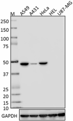
Whole cell lysates (15 µg protein) from A549, A431, HeLa, HE... 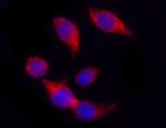
MCF-7 cell extract was resolved by electrophoresis, transfer... 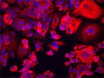
MCF-7 cells were stained with anti-Cytokeratin 18 (clone DA-... -
Alexa Fluor® 647 anti-Cytokeratin 18
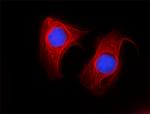
MCF-7 cells were fixed with 1% paraformaldehyde (PFA) for 10... -
Alexa Fluor® 594 anti-Cytokeratin 18
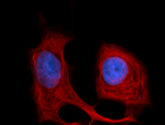
MCF-7 cells were fixed with 1% paraformaldehyde (PFA) for 10... 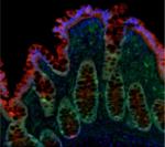
Human paraffin-embedded colon tissue was prepared with stand... -
Alexa Fluor® 488 anti-Cytokeratin 18
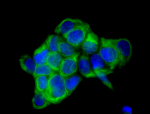
MCF-7 cells were fixed with 1% paraformaldehyde (PFA) for te... 
Multiplexed IHC staining of Alexa Fluor® 488 anti-CK18 (...

 Login / Register


Follow Us