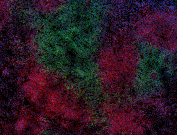
Paraformaldehyde-fixed (4%), 500 µm-thick mouse spleen section was processed according to the Ce3D™ Tissue Clearing Kit protocol (Cat. No. 480401). The section was anti-mouse CD172a (SIRPα) (clone P84) Alexa Fluor® 594 at 5 µg/mL (cyan), and anti-mouse CD200 (OX2) (clone OX-90) Alexa Fluor® 647 at 5 µlg/mL (magenta). The section was then optically cleared and mounted in a sample chamber. The image was captured with a 10X objective using Zeiss 780 confocal microscope and processed by Imaris image analysis software. Watch the video.
Paraformaldehyde-fixed (4%), 500 µm-thick mouse spleen section was processed according to the Ce3D™ Tissue Clearing Kit protocol (Cat. No. 480401). The section was anti-mouse CD172a (SIRPα) (clone P84) Alexa Fluor® 594 at 5 µg/mL (cyan), and anti-mouse CD200 (OX2) (clone OX-90) Alexa Fluor® 647 at 5 µlg/mL (magenta). The section was then optically cleared and mounted in a sample chamber. The image was captured with a 10X objective using Zeiss 780 confocal microscope and processed by Imaris image analysis software. Watch the video.
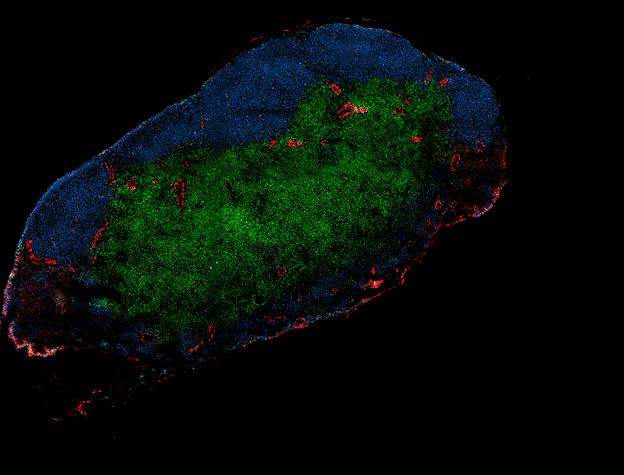
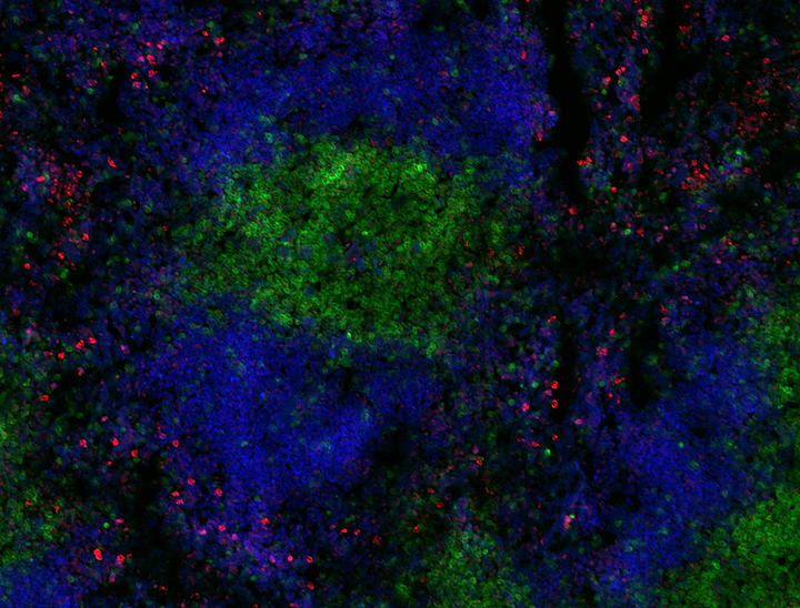
Paraformaldehyde-fixed (4%), 500 µm-thick mouse spleen section was processed according to the Ce3D™ Tissue Clearing Kit protocol (Cat. No. 480401). The section was costained with anti-mouse CD169 (Siglec-1) (clone 3D6.112) Alexa Fluor® 488 at 5 µg/mL (green), anti-mouse I-A/I-E (clone M5/114.15.2) Alexa Fluor® 594 at 5 µg/mL (blue), and anti-mouse CD3ε (clone KT3.1.1) Alexa Fluor® 647 at 5 µg/mL (magenta). The section was then optically cleared and mounted in a sample chamber. The image was captured with a 10X objective using Zeiss 780 confocal microscope and processed by Imaris image analysis software. Watch the video.
Paraformaldehyde-fixed (4%), mouse intestine section was processed according to the Ce3D™ Tissue Clearing Kit protocol (Cat. No. 480401). The section was costained with anti-Tubulin β 3 (TUBB3) (clone TUJ1) Alexa Fluor® 594 at 5 µg/mL (green), and anti-mouse CD45 (clone 30-F11) Alexa Fluor® 647 at 5 µg/mL (magenta). The section was then optically cleared and mounted in a sample chamber. The image was captured with a 20X objective using Zeiss 780 confocal microscope and processed by Imaris image analysis software. Watch the video.
Input string was not in a correct format.
Input string was not in a correct format.
Cat #
Size
Price
Quantity
Check Availability
Save
427709
50 mL
89 CHF
427710
250 mL
265 CHF
Description
Ce3D™ Wash Buffer is a ready-to-use buffer especially formulated for use as washing buffer with Ce3D™ tissue clearing reagent for 3D IHC application.

 Login / Register


Follow Us