- Clone
- P4G7 (See other available formats)
- Regulatory Status
- RUO
- Other Names
- Polyubiquitin-C, Polyubiquitin B, RPS 27A, RPS27A, UBA 52, UBA 80, UBA52, UBA80, UBB, UBC, UBCEP 1, UBCEP 2, UBCEP1, UBCEP2, Ubiquitin, ubiquitin B
- Isotype
- Mouse IgG1, κ
- Ave. Rating
- Submit a Review
- Product Citations
- publications
-

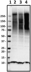
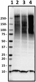
Western blot of HRP anti-Ubiquitin antibody (clone P4G7). Lane 1: Molecular weight marker; Lane 2: 30 µg of human brain lysate; Lane 3: 30 µg of mouse brain lysate; Lane 4: 30 µg of rat brain lysate. The blot was incubated with 2.5 µg/mL of the primary antibody overnight at 4°C. Enhanced chemiluminescence was used as the detection system (Cat. No. 426303). -

Western blot of HRP anti-Ubiquitin antibody (clone P4G7). Lane 1: Molecular weight marker; Lane 2: 20 µg of HepG2 cell lysate; Lane 3: 20 µg of NIH3T3 cell lysate. The blot was incubated with 5 µg/mL of the primary antibody overnight at 4°C. Enhanced chemiluminescence was used as the detection system (Cat. No. 426303). -

Western blot of HRP anti-Ubiquitin antibody (clone P4G7). Lane 1: Molecular weight marker; Lane 2: Drosophila head lysate (1 head). The blot was incubated with 10 µg/mL of the primary antibody overnight at 4°C. Enhanced chemiluminescence was used as the detection system (Cat. No. 426303). -

IHC staining of HRP anti-Ubiquitin antibody (clone P4G7) on formalin-fixed paraffin-embedded AD brain tissue. Following antigen retrieval using Sodium Citrate H.I.E.R, the tissue was incubated with 5 µg/ml of the primary antibody overnight at 4°C. DAB was used for detection followed by hematoxylin counterstaining, according to the protocol provided. The image was captured with a 40X objective. Scale bar: 50 µm -

IHC staining of HRP anti-Ubiquitin antibody (clone P4G7) on formalin-fixed paraffin-embedded AD brain tissue. Following antigen retrieval using Sodium Citrate H.I.E.R, the tissue was incubated with 5 µg/ml of the primary antibody overnight at 4°C. DAB was used for detection followed by hematoxylin counterstaining, according to the protocol provided. The image was captured with a 40X objective. Scale bar: 50 µm
| Cat # | Size | Price | Quantity Check Availability | Save | ||
|---|---|---|---|---|---|---|
| 838705 | 25 µg | 147 CHF | ||||
| 838706 | 100 µg | 311 CHF | ||||
Ubiquitin is a small (8.5 kD) regulatory protein that is ubiquitously expressed in tissues of eukaryotic organisms. There are four genes in the human genome that produce ubiquitin; UBB, UBC, UBA52 and RPS27A. UBA52 and RPS27A genes code for a single copy of ubiquitin fused to the ribosomal proteins L40 and S27a, respectively. The UBB and UBC genes code for polyubiquitin precursor proteins.
Ubiquitination is a post-translational modification where a ubiquitin subunit is attached to a protein. Addition of ubiquitin can signal for degradation via the proteasome, alter cellular location, promote or prevent protein interactions, or affect activity. Ubiquitination is carried out stepwise by ubiquitin-activating enzymes (E1s), ubiquitin-conjugating enzymes (E2s), and ubiquitin ligases (E3s), respectively. The cascade results in the binding of ubiquitin to lysine residues on the protein substrate via an isopeptide bond, cysteine residues through a thioester bond, serine and threonine residues through an ester bond, or the amino group of the protein's N-terminus via a peptide bond.
Proteins can be modified either by a single ubiquitin unit (monoubiquitination) or a chain of ubiquitin molecules (polyubiquitination). Proteins destined for degradation by the proteasome are usually ubiquitinated on lysine residues K48 and K29, while other polyubiquitinations (e.g. on K63, K11, K6) and monoubiquitinations may regulate processes such as endocytic trafficking, inflammation, translation and DNA repair.
A frameshift mutation in ubiquitin B can result in a truncated peptide missing the C-terminal glycine. This abnormal peptide, known as UBB+1, has been shown to accumulate selectively in Alzheimer's disease and other tauopathies.
Product Details
- Verified Reactivity
- Human, Mouse, Rat, Drosophila
- Antibody Type
- Monoclonal
- Host Species
- Mouse
- Immunogen
- Clone P4G7 was raised against denatured bovine ubiquitin and recognizes ubiquitin, polyubiquitin, and ubiquitin-conjugated proteins.
- Formulation
- This antibody is provided in 50% glycerol in aqueous buffered solutions with preservatives.
- Preparation
- The antibody was purified by affinity chromatography and conjugated with HRP under optimal conditions.
- Concentration
- 0.5 mg/ml
- Storage & Handling
- Upon receipt, the antibody solution should be stored undiluted at -20°C, and protected from prolonged exposure to light.
- Application
-
WB - Quality tested
IHC-P - Verified - Recommended Usage
-
Each lot of this antibody is quality control tested by Western blotting. For Western blotting, the suggested use of this reagent is 2.5 - 10 µg per ml. For immunohistochemistry on formalin-fixed paraffin-embedded tissue, a concentration range of 2.5 - 5.0 µg/ml is suggested. It is recommended that the reagent be titrated for optimal performance for each application.
- Application Notes
-
The ubiquitin protein is extremely well conserved and thus the antibody has extensive species cross-reactivity from yeast to human. This antibody was specifically developed to detect ubiquitin and ubiquitin-substrate conjugates by immunoblotting.
-
Application References
(PubMed link indicates BioLegend citation) - RRID
-
AB_2801179 (BioLegend Cat. No. 838705)
AB_2801180 (BioLegend Cat. No. 838706)
Antigen Details
- Structure
- Ubiquitin is a 76 amino acid protein with a molecular mass of 8.5 kD.
- Distribution
-
Found in almost all tissues of eukaryotic organisms
- Function
- Regulatory protein
- Biology Area
- Cell Biology, Neurodegeneration, Neuroscience, Neuroscience Cell Markers, Protein Trafficking and Clearance, Signal Transduction
- Molecular Family
- Autophagosome Markers
- Antigen References
-
1. Hatanaka H, et al. 2009. J. Biol. Chem. 23:15448. (WB) PubMed
2. Sarkari F, et al. 2013. J. Biol. Chem. 23:16975. (WB) PubMed - Gene ID
- 7316 View all products for this Gene ID
- UniProt
- View information about Ubiquitin on UniProt.org
Related FAQs
Other Formats
View All Ubiquitin Reagents Request Custom Conjugation| Description | Clone | Applications |
|---|---|---|
| Purified anti-Ubiquitin | P4G7 | WB,IHC-P |
| HRP anti-Ubiquitin | P4G7 | WB,IHC-P |
| Biotin anti-Ubiquitin | P4G7 | IHC-P |
| Alexa Fluor® 647 anti-Ubiquitin | P4G7 | IHC-P |
| Alexa Fluor® 488 anti-Ubiquitin | P4G7 | IHC-P |
Compare Data Across All Formats
This data display is provided for general comparisons between formats.
Your actual data may vary due to variations in samples, target cells, instruments and their settings, staining conditions, and other factors.
If you need assistance with selecting the best format contact our expert technical support team.
-
Purified anti-Ubiquitin
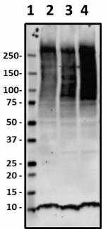
Western blot of purified anti-Ubiquitin antibody (clone P4G7... 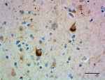
IHC staining of purified anti-Ubiquitin antibody (clone P4G7... 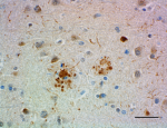
IHC staining of purified anti-Ubiquitin antibody (clone P4G7... 
Western blot of purified anti-Ubiquitin antibody (clone P4G7... 
Western blot of purified anti-Ubiquitin antibody (clone P4G7... -
HRP anti-Ubiquitin
Western blot of HRP anti-Ubiquitin antibody (clone P4G7). La... 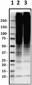
Western blot of HRP anti-Ubiquitin antibody (clone P4G7). La... 
Western blot of HRP anti-Ubiquitin antibody (clone P4G7). La... 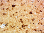
IHC staining of HRP anti-Ubiquitin antibody (clone P4G7) on ... 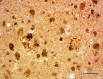
IHC staining of HRP anti-Ubiquitin antibody (clone P4G7) on ... -
Biotin anti-Ubiquitin
IHC staining of Biotin anti-Ubiquitin antibody (clone P4G7) ... 
IHC staining of Biotin anti-Ubiquitin antibody (clone P4G7) ... 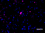
IHC staining of Biotin anti-Ubiquitin antibody (clone P4G7) ... -
Alexa Fluor® 647 anti-Ubiquitin
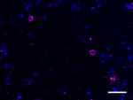
IHC staining of Alexa Fluor® 647 anti-Ubiquitin antibody (cl... 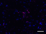
IHC staining of Alexa Fluor® 647 anti-Ubiquitin antibody (cl... 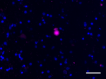
IHC staining of Alexa Fluor® 647 anti-Ubiquitin antibody (cl... -
Alexa Fluor® 488 anti-Ubiquitin
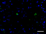
IHC staining of Alexa Fluor® 488 anti-Ubiquitin antibody (cl... 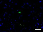
IHC staining of Alexa Fluor® 488 anti-Ubiquitin antibody (cl...
 Login / Register


Follow Us