
 Login / Register


- Clone
- Poly19056 (See other available formats)
- Regulatory Status
- RUO
- Other Names
- Keratin, type II cytoskeletal 1, K1, CK-1, keratin 86, cytokeratin-1, 67 kD cytokeratin, type-II keratin Kb1, keratin complex 2, basic, gene 1
- Previously
-
Covance Catalog# PRB-165P
- Isotype
- Rabbit Polyclonal IgG
- Ave. Rating
- Submit a Review
- Product Citations
- 21 publications


| Cat # | Size | Price | Quantity Check Availability | Save | ||
|---|---|---|---|---|---|---|
| 905602 | 25 µL | 72€ | ||||
| 905601 | 100 µL | 231€ | ||||
Keratin 1 belongs to the keratin family of proteins which are fibrous structural proteins. It is a type II cytokeratin that forms the major cytoskeleton in suprabasal keratinocytes along with heterodimer type I partner KRT10 form. KRT1 and KRT10 mutations lead to congenital epidermolytic ichthyosis characterized by skin erosions and hyperkeratosis.
Product DetailsProduct Details
- Verified Reactivity
- Mouse, Rat
- Antibody Type
- Polyclonal
- Host Species
- Rabbit
- Immunogen
- This monospecific polyclonal antibody was raised against a peptide sequence derived from the C-terminus of the mouse keratin 1 protein.
- Formulation
- Phosphate-buffered solution + 0.03% Thimerosal.
- Preparation
- The antibody was purified by affinity chromatography.
- Concentration
- 1.0 mg/mL
- Storage & Handling
- The antibody solution should be stored undiluted between 2°C and 8°C. Please note the storage condition for this antibody has been changed from -20°C to between 2°C and 8°C. You can also check your vial or your CoA to find the most accurate storage condition for this antibody.
- Application
-
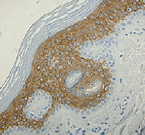
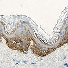
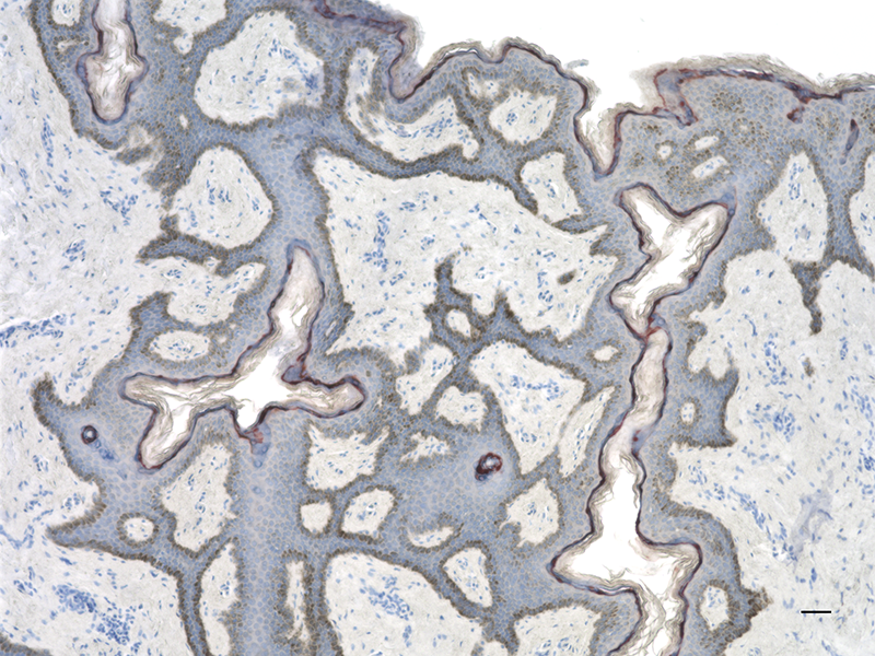
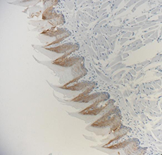
IHC-P - Quality tested
WB - Verified
IHC-F - Reported in the literature, not tested in house - Recommended Usage
-
Each lot of this antibody is quality control tested by formalin-fixed paraffin-embedded immunohistochemical staining. For immunohistochemistry, a dilution of 1:500 is suggested. For Western blotting, the suggested use of this reagent is 1.0 µg per mL (1:1000 dilution). For immunocytochemistry, a dilution of 1:500 is recommended. It is recommended that the reagent be titrated for optimal performance for each application.
- Application Notes
-
This antibody is effective in immunoblotting (WB), immunofluorescence (IF) and immunohistochemistry (IHC). This antibody has been successfully used in both frozen and paraffin embedded tissues.
*Predicted MW = 67 kD
This product may contain other non-IgG subtypes. - Application References
-
- Croyle MJ, et al. 2011. Development 138: 1675. (IHC, IF) PubMed
- Wirt SE, et al. 2010. J. Cell Biol. 191: 809. (IHC) PubMed
- Hu Y, et al. 2001. Nature. 410:710.
- Yuspa SH, et al. 1989. J Cell Biol. 109:1207.
- Roop DR, et al. 1984. J Biol Chem. 259:8037.
- Mailleux AA, et al. 2007. Dec Cell. 12:221. (IHC)
- IShitsuka Y, et al. 2013. J Invest Dermatol. 133:2566. (WB)
- Sengupta A, et al. 2010. PLoS One. 5:12249. (IHC) PubMed
- Suzuki K, et al. 2009. Development. 136:367. (IHC) PubMed
- Levy V, et al. 2007. FASEB J. 21:1358. (IHC) PubMed
- Osakarsson T, et al. 2006. Genes Dev. 20:2024. (IHC) PubMed
- Liang Y. 2011. Patholog Res Int. 2011:93674. (IHC) PubMed
- Sachs N, et al. 2012. PNAS. 109:21468. (IF)
- Vidal VP, et al. 2005. Curr Biol. 15:1340. (IHC)
- Product Citations
-
- RRID
-
AB_2801250 (BioLegend Cat. No. 905602)
AB_2565051 (BioLegend Cat. No. 905601)
Antigen Details
- Biology Area
- Cell Biology, Neuroscience, Neuroscience Cell Markers
- Molecular Family
- Intermediate Filaments
- Gene ID
- 16678 View all products for this Gene ID
- UniProt
- View information about Keratin 1 on UniProt.org

Related FAQs
Other Formats
View All Keratin 1 Reagents Request Custom Conjugation| Description | Clone | Applications |
|---|---|---|
| Purified anti-Keratin 1 | Poly19056 | IHC-P,WB,IHC-F |
Customers Also Purchased
Compare Data Across All Formats
This data display is provided for general comparisons between formats.
Your actual data may vary due to variations in samples, target cells, instruments and their settings, staining conditions, and other factors.
If you need assistance with selecting the best format contact our expert technical support team.


Follow Us