


- Clone
- A19290E (See other available formats)
- Regulatory Status
- RUO
- Other Names
- IBA1, Allograft Inflammatory Factor 1, IRT-1, Interferon Gamma Responsive Transcript
- Isotype
- Mouse IgG2a, κ
- Ave. Rating
- Submit a Review


| Cat # | Size | Price | Save |
|---|---|---|---|
| 615351 | 25 µg | ¥26,400 | |
| 615352 | 100 µg | ¥86,900 |
As a cytoplasmic scaffold protein, IBA1 contains Ca2+ binding EF-hand and PDZ interaction domains, which are important for mediating intracellular signaling complexes. IBA1, also known as AIF-1, was first observed in rodent microglial cells and has been shown to stain cells in both normal and pathological lesions in rodents. In addition to the central nervous system, IBA1 has also been shown to stain in macrophage subpopulations. In macrophages, expression of IBA1 promotes pro-inflammatory responses. In microglial cells, IBA1 is important in the modulation of synaptic activities and is upregulated in response to nerve damage.
Product DetailsProduct Details
- Verified Reactivity
- Human
- Antibody Type
- Monoclonal
- Host Species
- Mouse
- Immunogen
- Synthetic peptide from human IBA1
- Formulation
- Phosphate-buffered solution, pH 7.2, containing 0.09% sodium azide
- Preparation
- The antibody was purified by affinity chromatography.
- Concentration
- 0.5 mg/mL
- Storage & Handling
- The antibody solution should be stored undiluted between 2°C and 8°C.
- Application
-
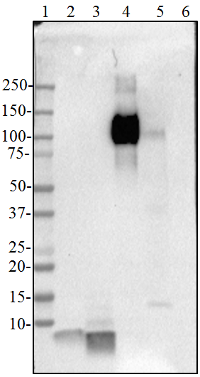
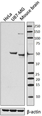
WB - Quality tested
IHC-P, ICFC - Verified - Recommended Usage
-
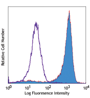
Each lot of this antibody is quality control tested by western blotting. For western blotting, the suggested use of this reagent is 1.0 µg/mL. For immunohistochemistry on formalin-fixed paraffin-embedded tissue sections, a concentration range of 1.0 - 10.0 µg/mL is suggested. For flow cytometric staining, the suggested use of this reagent is ≤ 0.125 µg per million cells in 100 µL volume. It is recommended that the reagent be titrated for optimal performance for each application.
- Application Notes
-
This clone is not mouse or rat reactive.
This clone is not suitable for ICC. - RRID
-
AB_3083402 (BioLegend Cat. No. 615351)
AB_3083402 (BioLegend Cat. No. 615352)
Antigen Details
- Structure
- IBA1 is a 147 amino acid protein with a predicted molecular weight of 17 kD.
- Distribution
-
Expressed in microglia and macrophages
- Function
- Plays a role in inflammatory response and modulates synaptic activities
- Interaction
- Interacts with LCP1
- Ligand/Receptor
- Calcium
- Cell Type
- Lymphocytes, Macrophages, Microglia, T cells
- Biology Area
- Apoptosis/Tumor Suppressors/Cell Death, Cell Death, Immunology, Innate Immunity, Neuroinflammation, Neuroscience, Neuroscience Cell Markers, Synaptic Biology
- Molecular Family
- Innate Immune Signaling
- Antigen References
-
- Elizondo D. M. et al. 2017. Front Immunol. 8:1502.
- Mikkelsen HB, et al. 2017. Anat Rec (Hoboken). 300:1114-1122.
- Shapiro LA, et al. 2009. Brain Res. 1266:29-36.
- Gene ID
- 199 View all products for this Gene ID
- UniProt
- View information about Iba1 on UniProt.org

Related FAQs
Other Formats
View All Iba1 Reagents Request Custom Conjugation| Description | Clone | Applications |
|---|---|---|
| Purified anti-IBA1 | A19290E | WB,IHC-P,ICFC |
Customers Also Purchased

Compare Data Across All Formats
This data display is provided for general comparisons between formats.
Your actual data may vary due to variations in samples, target cells, instruments and their settings, staining conditions, and other factors.
If you need assistance with selecting the best format contact our expert technical support team.


Follow Us