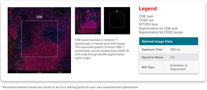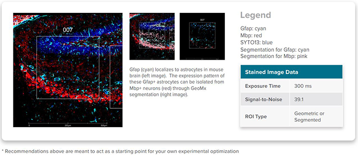
A robust method of selecting such ROIs involves the use of our fluor-conjugated antibodies as morphology markers in tissue samples to label tissue compartments and cell types. The morphology of the sample then guides the selection of ROIs based on biologically relevant features. Using the integrated segmentation algorithm in DSP software, ROIs may be automatically divided into discrete biological compartments or areas of illumination (AOI) for precise profiling and used to answer specific biological questions.
Image from NanoString Morphology Marker datasheet on CD8a in cytotoxic T lymphocytes from FFPE human tonsil tissue.
Image from NanoString Morphology Marker datasheet on GFAP (Glial Fibrillary Acidic Protein) in mature astrocytes from fresh frozen mouse brain tissue.
See the table below for a current list of verified GeoMx® DSP morphology markers for FFPE, fresh frozen, or fixed frozen tissues.
View the full list of BioLegend’s fluor-conjugated antibodies verified as morphology markers.
|
Species |
# of Available Markers |
Fluorophore |
Tissue Type |
GeoMx® DSP Assay Supported |
Representative Datasheets |
|
Human (Qualified1 or Verified2) |
14 |
Alexa Fluor® (various), PE |
FFPE, FF |
RNA |
|
|
Mouse (Qualified1 or Verified2) |
5 |
Alexa Fluor® 647 |
FFPE, FF |
RNA |
|
|
Human (TAP3) |
4 |
Alexa Fluor® (various), Unconjugated antibodies (detected with secondary) |
FFPE |
RNA and Protein |
--- |
|
Mouse (TAP3) |
4 |
Alexa Fluor® (various), Unconjugated antibodies (detected with secondary) |
FFPE |
RNA and Protein |
--- |
- Verified markers have gone through extensive testing on multiple tissues, when possible, with target-specific positive tissue staining verified by an experienced pathologist.
- Qualified markers have been shown by NanoString to work in at least one tissue sample.
- The GeoMx® DSP Technology Access Program (TAP) team has verified the marker in at least one customer-provided tissue sample.
 Login / Register


Follow Us