- Clone
- Poly19053 (See other available formats)
- Regulatory Status
- IVD
- Isotype
- Rabbit Polyclonal
| Cat # | Size | Price | Quantity Check Availability | Save | ||
|---|---|---|---|---|---|---|
| 905301 | 100 µL | 352€ | ||||
This antibody is effective in immunohistochemistry (IHC).
This product may contain other non-IgG subtypes.
Product Details
- Product Information
-
Intended Use:
In Vitro Diagnostic (IVD). Use in immunohistochemistry (IHC) test methods only.
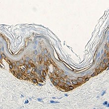
The polyclonal antibody Poly19053 is used for the in vitro examination of frozen or paraffin-embedded human skin tissue sections using immunohistochemistry (IHC) methods for the qualitative identification of Keratin 14. The clinical interpretation of any staining or its absence should be complemented by morphological studies and proper controls and should be evaluated within the context of the patient's clinical history and other diagnostic tests by a qualified pathologist.
- Reactivity
- Human
- Formulation
- Phosphate-buffered solution + 0.03% Thimerosal.
- Preparation
- The antibody was purified by peptide affinity chromatography.
- Concentration
- 1.0 mg/mL
- Storage & Handling
- When stored at ≤ -20°C, product is stable until the date shown on the label. Avoid repeated freeze-thaw cycles to prevent denaturing the antibody. If thawed and stored between 2°C and 8°C, product is stable for 24 months from the date of thaw or until the expiry date on the label, whichever comes first.
- Recommended Usage
-
Each lot of this antibody is quality control tested by immunohistochemical staining of formalin-fixed parafin-embedded sections of normal human skin. Frozen human skin tisue has been veified during product development
The optimal working dilution should be determined for each specific assay condition.
• IHC: 1:1,000 with either biotin based detection systems such as USA Ultra Streptavidin Detection (Cat. No. 929501).
Tissue Sections: Paraffin-embedded tissues, frozen tissues
Pretreatment: For optimal staining, the sections should be pretreated with an antigen unmasking solution such as Citrate Buffer Retrieval solution (Cat. No. 928502).
Incubation: 60 minutes at room temperature - Application References
-
- Wend P, et al. Wnt/?-catenin signalling induces MLL to create epigenetic changes in salivary gland tumours. EMBO J., Jun 2013.
- Welm AL, Kim S, Welm BE, Bishop JM. MET and MYC cooperate in mammary tumorigenesis. Proc Natl Acad Sci USA 102(12):4324-9, 2005. [IHC] PubMed
- Hu Y, Baud V, Oga T, Kim KI, Yoshida K, Karin M. IKKalpha controls formation of the epidermis independently of NF-kappaB. Nature 410:710-714, 2001.
- Yuspa SH, Kilkenny AD, Steinert PM, Roop DR. Expression of murine epidermal differentiation markers is tightly regulated by restricted extracellular calcium concentrations in-vitro. J Cell Biol 109:1207-1217, 1989.
- Roop DR, Cheng CK, Titterington L, Meyers CA, Stanley JR, Steinert PM, Yuspa SH. Synthetic peptides corresponding to keratin subunits elicit highly specific antibodies. J Biol Chem 259:8037-8040, 1984.
- Liang Y. 2011. Patholog Res Int. 2011:93674. (IHC) PubMed
- Easter SL, et al. 2014. PLoS One. 9:113247. (IF, WB) PubMed
- Frew IJ, et al. 2008. Mol Cell Biol.28:4536. (IHC) PubMed
- Levy V, et al. 2007. FASEB J. 21:1358. (IHC) PubMed
- Sengupta A, et al. 2010. PLoS One. 5:12249. (IHC) PubMed
- Smith BA, et al. 2012. Genes Cancer.3:550. (IHC) PubMed
- RRID
-
AB_2565048 (BioLegend Cat. No. 905301)
- Disclaimer
-
WARNINGS AND PRECAUTIONS
- Use appropriate personal protective equipment and safety practices per universal precautions when working with this reagent. Refer to the reagent safety data sheet.
- All specimens, samples and any material coming in contact with them should be considered potentially infectious and should be disposed of with proper precautions and in accordance with federal, state and local regulations.
- Do not use this reagent beyond the expiration date stated on the label.
- Do not use this reagent if it appears cloudy or if there is any change in the appearance of the reagent as these may be indication of possible deterioration.
Related FAQs
Customers Also Purchased
Compare Data Across All Formats
This data display is provided for general comparisons between formats.
Your actual data may vary due to variations in samples, target cells, instruments and their settings, staining conditions, and other factors.
If you need assistance with selecting the best format contact our expert technical support team.
-
Keratin 14 Polyclonal Antibody, Purified
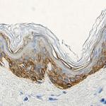
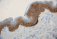
Strong cytoplasmic staining of the epidermis in normal human... 

-
Purified anti-Keratin 14
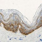
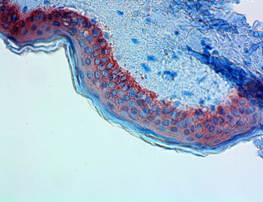
IHC staining of anti-Keratin 14 antibody (Poly19053) on form... 
Western blot of anti-Keratin 14 antibody (Poly19053). Lane 1... 
Western blot analysis of cell lysates from HaCaT (Positive c... 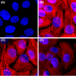
Immunofluorescence of HaCaT cells with (A) Rabbit Isotype co... 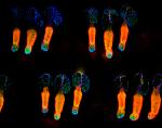
Whole mount mouse tail epidermis was stained with purified a...

 Login / Register




Follow Us