Purified anti-Keratin 14 Antibody

Pricing & Availability
Clone
Poly19053 (See other available formats)
Regulatory Status
RUO
Other Names
Keratin, type I cytoskeletal 14, CK-14, cytokeratin 14, keratin 14 (epidermolysis bullosa simplex, Dowling-Meara, Koebner)
Isotype
Rabbit Polyclonal
Ave. Rating
Submit a Review
Product Citations
publications
1_Poly19053_PURE_Keratin14_Antibody_1_WB_102317
Western blot analysis of cell lysates from HaCaT (Positive control) and U87-MG (Low expression negative control) cells using Keratin 14 Rabbit primary antibody and HRP Donkey anti-Rabbit secondary antibody (#406401). Direct-Blot™ HRP anti-β-actin (#643807) was used as a loading control.
  • 1_Poly19053_PURE_Keratin14_Antibody_1_WB_102317
    Western blot analysis of cell lysates from HaCaT (Positive control) and U87-MG (Low expression negative control) cells using Keratin 14 Rabbit primary antibody and HRP Donkey anti-Rabbit secondary antibody (#406401). Direct-Blot™ HRP anti-β-actin (#643807) was used as a loading control.
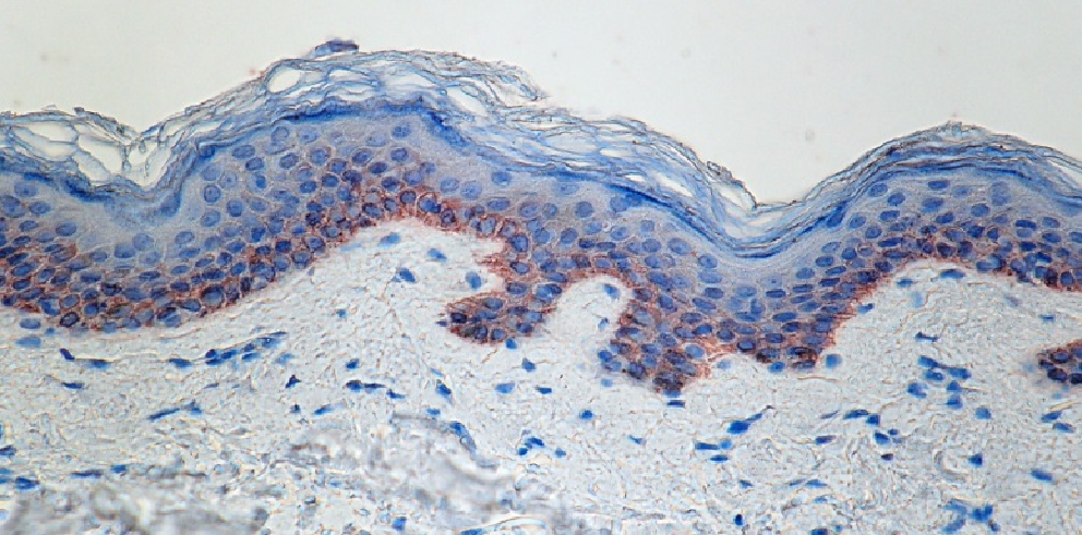
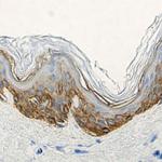
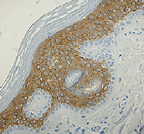
  • 2_Poly19053_PURE_Keratin14_Antibody_1_IHC_052716
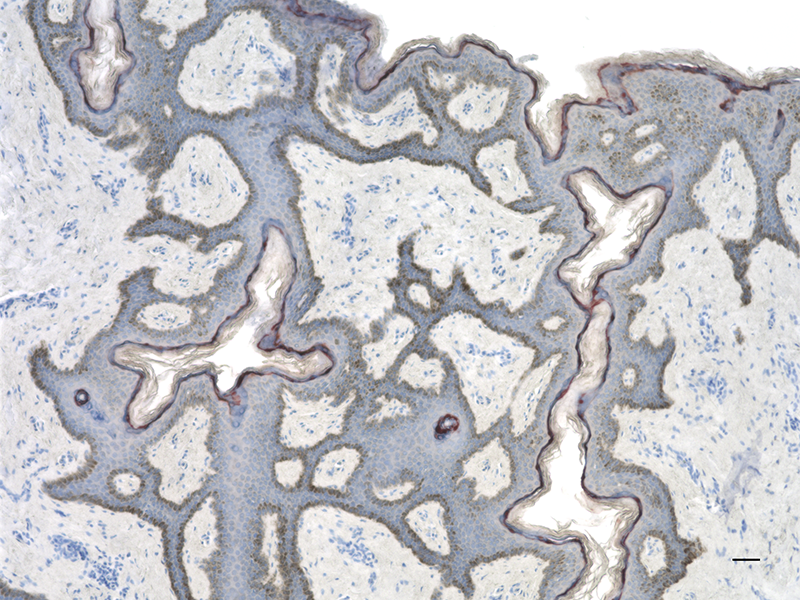
    IHC staining of anti-Keratin 14 antibody (Poly19053) on formalin-fixed paraffin-embedded normal human skin. After antigen retrieval using Sodium Citrate, the tissue was incubated with the primary antibody at 1 µg/ml for 60 minutes at room temperature. BioLegend’s Ultra Streptavidin (USA) HRP Detection Kit was used for detection followed by hematoxylin counterstaining, according to the protocol provided.
  • 3_Poly19053_PURE_Keratin14_Antibody_2_WB_052716
    Western blot of anti-Keratin 14 antibody (Poly19053). Lane 1: Molecular weight marker; Lane 2: Human skin lysate. The blot was incubated with the primary antibody at 1 µg/ml, overnight at 4°C, followed by incubation with a fluorescent labeled goat anti-mouse secondary antibody.
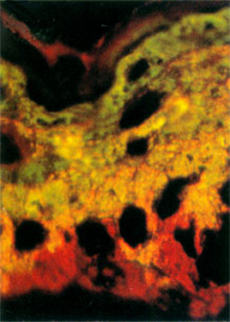
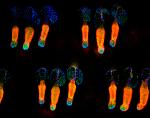
  • KifLiakath
    Whole mount mouse tail epidermis was stained with purified anti-Keratin 14 (green, clone Poly19053), purified anti-Keratin 15 (orange, clone Poly18339) and DAPI (blue). Image generously submitted to the 2017 Cell Life Imaging Competition by Kif Liakath.
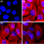
  • Poly19053_PURE_Keratin-14_Antibody_ICC_112817
    Immunofluorescence of HaCaT cells with (A) Rabbit Isotype control (Cat. No. 910801) or (B-D) Keratin 14 primary Antibody (Clone Poly 19053). 2 µg/ml (1:250 dilution) Alexa Fluor® 594 (red) Donkey anti-Rabbit IgG (Cat. No. 406418) was used as secondary antibody. Nuclei were counterstained with DAPI (blue). The image was captured with a 60X objective using KEYENCE BZ-X700 fluorescence microscope. Exposure time (Seconds) for (A) is 1/30, and (B-D) is 1/60. Anti-Keratin 14 primary antibody concentrations for (A, B) is 2 µg/ml (1:250 dilution), (C) is 1 µg/ml (1:1000 dilution), (D) is 0.5 µg/ml (1:1000 dilution).
See high resolution IHC data...
Cat # Size Price Quantity Check Availability Save
905303 25 µg 100€
Check Availability


Need larger quantities of this item?
Request Bulk Quote
905304 100 µg 280€
Check Availability


Need larger quantities of this item?
Request Bulk Quote
Description

Keratin 14 is a member of the keratin family, the most diverse group of intermediate filaments. This gene product, a type I keratin, is usually found as a heterotetramer with two keratin 5 molecules, a type II keratin. Together they form the cytoskeleton of epithelial cells. Mutations in the genes for these keratins are associated with epidermolysis bullosa simplex.

Product Details
Technical Data Sheet (pdf)

Product Details

Verified Reactivity
Human, Mouse, Rat
Reported Reactivity
Dog, Non-Human Primate
Antibody Type
Polyclonal
Host Species
Rabbit
Formulation
Phosphate-buffered solution, pH 7.2, containing 0.09% sodium azide.
Preparation
The antibody was purified by affinity chromatography.
Storage & Handling
The antibody solution should be stored undiluted between 2°C and 8°C.
Application

IHC-P - Quality tested
ICC, WB - Verified

Recommended Usage

Each lot of this antibody is quality control tested by formalin-fixed paraffin-embedded immunohistochemical staining. For immunohistochemistry, a concentration range of 0.1 - 10 μg/ml is suggested. For Western blotting, a concentration range of 0.5 - 2 ug/ml (1:250-1:1000) is suggested. For immunocytochemistry, a concentration range of 0.5 - 2.0 μg/ml (1:250 - 1:1000) is recommended. It is recommended that the reagent be titrated for optimal performance for each application.

Application Notes


This product may contain other non-IgG subtypes.

Application References
  1. Hu Y, et al. 2001. Nature 410:710. PubMed
  2. Yuspa SH, et al. 1989. J. Cell. Biol. 109:1207. PubMed
  3.  
Product Citations
  1. Miyauchi K, et al. 2021. Front Immunol. 12:663177. PubMed
  2. Sipilä K, et al. 2022. Dev Cell. 57:1453. PubMed
  3. Raja E, et al. 2022. EMBO Rep. 23:e55478. PubMed
  4. Das A, et al. 2022. J Invest Dermatol. 142:679. PubMed
  5. Yuan X, et al. 2023. J Periodontol. 94:277. PubMed
  6. Feigin CY, et al. 2023. Sci Adv. 9:eade7511. PubMed
  7. Gao J, et al. 2023. Sci Rep. 13:5144. PubMed
  8. Okada N, et al. 2023. Commun Biol. 6:596. PubMed
  9. Fu R, et al. 2019. Nat Commun. 10:3210. PubMed
  10. Changarathil G, et al. 2019. PLoS One. 14:e0215908. PubMed
  11. Abbasi S, et al. 2020. Cell Stem Cell. 27(3):396-412.e6. PubMed
  12. Moehring F et al. 2018. eLife. 7 pii: e31684. PubMed
  13. Yang K, et al. 2022. J Clin Invest. 132:. PubMed
  14. Guo Q, et al. 2022. Nat Commun. 13:3837. PubMed
  15. Yip HYK, et al. 2021. STAR Protocols. 2(3):100765. PubMed
  16. Altmeier S, et al. 2016. PLoS Pathog. 12: 1005882. PubMed
  17. Wang R, et al. 2021. Front Cell Dev Biol. 9:772669. PubMed
  18. Ishii R, et al. 2020. Development. . PubMed
  19. Jones KB et al. 2018. Cell stem cell. 24(1):183-192 . PubMed
  20. Adams CR, et al. 2019. Elife. 8. PubMed
  21. Reehorst CM, et al. 2021. Development. 148:. PubMed
  22. Johansson E, et al. 2021. Sci Rep. 11:22329. PubMed
  23. Yip HYK, et al. 2020. Molecular Cell. 80(2):279-295.e8. PubMed
  24. Nishiguchi MA et al. 2018. Cell reports. 24(13):3383-3392 . PubMed
  25. Pruett L, et al. 2021. J Mater Chem B. 9:7132. PubMed
  26. Christin JR, et al. 2020. Cell Reports. 31(10):107742. PubMed
  27. Koenig U, et al. 2019. Autophagy. :1. PubMed
  28. Cowie AM et al. 2018. The Journal of Neuroscience. 38(25):5807-5825 . PubMed
  29. Zhong Z, et al. 2021. Biomaterials. 267:120462. PubMed
  30. Ishii R, et al. 2020. Development. . PubMed
  31. Giraddi RR et al. 2018. Cell reports. 24(6):1653-1666 . PubMed
  32. Anderton H, et al. 2018. Cell Death Differ. 26:877. PubMed
  33. Anderton H, et al. 2017. J Invest Dermatol. 137:2371. PubMed
RRID
AB_2616896 (BioLegend Cat. No. 905303)
AB_2616896 (BioLegend Cat. No. 905304)

Antigen Details

Structure
Keratin 14 is a 472 amino acid protein with a molecular mass of 52 kD.
Distribution

Tissue distribution: skin.
Cellular distribution: extracellular, nucleus, cytoskeleton, and cytosol.

Function
Nonhelical tail domain is involved in promoting KRT5-KRT14 filaments to self-organize into large bundles, enhances the mechanical properties involved in resilience of keratin intermediate filaments in vitro.
Biology Area
Cell Biology, Cell Motility/Cytoskeleton/Structure, Neuroscience, Neuroscience Cell Markers
Molecular Family
Intermediate Filaments
Antigen References

1. Wend P, et al. 2013. EMBO J. 32(14):1977-89. PubMed
2. Welm AL, et al. 2005. Proc. Natl. Acad. Sci. USA 102(12):4324. (IHC) PubMed
3. Liang Y. 2011. Pathalog. Res. Int. 2011:93674. (IHC) PubMed

Gene ID
3861 View all products for this Gene ID
UniProt
View information about Keratin 14 on UniProt.org
Go To Top Version: 6    Revision Date: 08-12-2022

For Research Use Only. Not for diagnostic or therapeutic use.


This product is supplied subject to the terms and conditions, including the limited license, located at www.biolegend.com/terms) ("Terms") and may be used only as provided in the Terms. Without limiting the foregoing, BioLegend products may not be used for any Commercial Purpose as defined in the Terms, resold in any form, used in manufacturing, or reverse engineered, sequenced, or otherwise studied or used to learn its design or composition without express written approval of BioLegend. Regardless of the information given in this document, user is solely responsible for determining any license requirements necessary for user’s intended use and assumes all risk and liability arising from use of the product. BioLegend is not responsible for patent infringement or any other risks or liabilities whatsoever resulting from the use of its products.


BioLegend, the BioLegend logo, and all other trademarks are property of BioLegend, Inc. or their respective owners, and all rights are reserved.


8999 BioLegend Way, San Diego, CA 92121 www.biolegend.com
Toll-Free Phone: 1-877-Bio-Legend (246-5343) Phone: (858) 768-5800 Fax: (877) 455-9587

This data display is provided for general comparisons between formats.
Your actual data may vary due to variations in samples, target cells, instruments and their settings, staining conditions, and other factors.
If you need assistance with selecting the best format contact our expert technical support team.

ProductsHere

Login / Register
Remember me
Forgot your password? Reset password?
Create an Account