- Other Names
- Colony Stimulating Factor 1, Colony Stimulating Factor 1 (Macrophage), Macrophage Colony Stimulating Factor 1, CSF-1, MCSF, CSF1
- Ave. Rating
- Submit a Review
- Product Citations
- publications

-

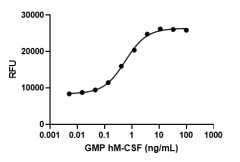
GMP recombinant human M-CSF induces proliferation of M-NFS-60 mouse myelogenous leukemia lymphoblast cells in a dose-dependent manner with an ED50 range of 0.5 - 2.0 ng/mL.
| Cat # | Size | Price | Quantity Check Availability | Save | ||
|---|---|---|---|---|---|---|
| 574814 | 25 µg | $347 | ||||
| 574816 | 100 µg | $1085 | ||||
Macrophage colony formation from precursors in murine bone marrow cultures. M-CSF is constitutively present at biologically active concentrations in human serum. It binds CD14+ monocytes and promotes the survival/proliferation of human peripheral blood monocytes. In addition, M-CSF enhances inducible monocyte functions including phagocytic activity, microbial killing, cytotoxicity for tumor cells as well as synthesis of inflammatory cytokines such as IL-1, TNF-α, and INF-γ in monocytes. M-CSF induces RANKL production in mature human osteoclasts; consequently, M-CSF is a potent stimulator of mature osteoclast resorbing activity. Also, M-CSF induces VEGF in human monocytes in human tumors; high levels of M-CSF, mononuclear phagocytes, and VEGF are associated with poor prognosis in patients with cancer. High levels of M-CSF have been associated with different pathologies such as pulmonary fibrosis and atherosclerosis. M-CSF binds to its receptor M-CSFR, and this receptor is shared by a second ligand, IL-34. Human M-CSF and IL-34 exhibit cross-species specificity – both bind to human and mouse M-CSF receptors.
Product DetailsProduct Details
- Source
- Human M-CSF, amino acids Glu33-Ser190 (Accession# NM_172212.2) was expressed in 293E cells.
- Molecular Mass
- The 179 amino acid recombinant protein has a predicted molecular mass of approximately 20.6 kD. The DTT-reduced and non-reduced protein migrate at approximately 25 - 35 kD and 55 - 70 kD respectively by SDS-PAGE. The N-terminal contains a His9-(SGGG)2-IEGR-tag.
- Purity
- > 95%, as determined by Coomassie stained SDS-PAGE
- Formulation
- 0.1 µm filtered protein solution is in PBS.
- Endotoxin Level
- Less than 0.1 EU per µg protein as determined by the LAL method
- Concentration
- 500 µg/mL
- Storage & Handling
- Unopened vial can be stored between 2°C and 8°C for up to 2 weeks, at -20°C for up to six months, or at -70°C or colder until the expiration date. For maximum results, quick spin vial prior to opening. The protein can be aliquoted and stored at -20°C or colder. Stock solutions can also be prepared at 50 - 100 µg/mL in appropriate sterile buffer, carrier protein such as 0.2 - 1% endotoxin-free BSA or HSA can be added when preparing the stock solution. Aliquots can be stored between 2°C and 8°C for up to one week or stored at -20°C or colder for up to 3 months. Avoid repeated freeze/thaw cycles.
- Activity
- ED50 = 0.5 - 2.0 ng/mL as measured by its ability to induce proliferation of M-NFS-60 mouse myelogenous leukemia lymphoblast cells.
- Application
-
Bioassay
Cell cultures - Application Notes
-
BioLegend carrier-free recombinant proteins provided in liquid format are shipped on blue ice. Our comparison testing data indicates that when handled and stored as recommended, the liquid format has equal or better stability and shelf-life compared to commercially available lyophilized proteins after reconstitution. Our liquid proteins are verified in-house to maintain activity after shipping on blue ice and are backed by our 100% satisfaction guarantee. If you have any concerns, contact us at tech@biolegend.com.
- Disclaimer
-
GMP Recombinant Proteins. BioLegend GMP recombinant proteins are manufactured in a dedicated GMP facility and compliant with ISO 13485:2016. For research or ex vivo cell processing use. Not for use in diagnostic or therapeutic procedures. Our processes include:
- Batch-to-batch consistency
- Material traceability
- Documented procedures
- Documented employee training
- Equipment maintenance and monitoring records
- Lot-specific certificates of analysis
- Quality audits per ISO 13485:2016
- QA review of released products
BioLegend GMP recombinant proteins are manufactured and tested in accordance with USP Chapter 1043, Ancillary Materials for Cell, Gene and Tissue-Engineered Products and Ph. Eur. Chapter 5.2.12.
Antigen Details
- Structure
- Disulfide-linked homodimer
- Distribution
-
Hematopoietic stem and progenitors, embryonic stem cells
- Function
- Cytokine that plays an essential role in the regulation of survival, proliferation and differentiation of hematopoietic precursor cells, especially mononuclear phagocytes, such as macrophages and monocytes.
- Interaction
- Monocytes, macrophages, mononuclear phagocyte precursors, microglia, proliferating smooth muscle cells, umbilical vein endothelial cells, and breast cancer cell lines
- Ligand/Receptor
- M-CSFR or CSF1R (CD115)
- Bioactivity
- Measured by its ability to induce proliferation of M-NFS-60 cells.
- Cell Type
- Embryonic Stem Cells, Hematopoietic stem and progenitors
- Biology Area
- Cell Biology, Cell Proliferation and Viability, Immunology, Stem Cells
- Molecular Family
- Cytokine/Chemokine Receptors, Growth Factors
- Antigen References
-
- Kawasaki ES, et al. 1985. Science 230:291.
- Wei S, et al. 2010. J. Leukoc. Biol. 88:495.
- Hodge JM, et al. 2011. PloS One 6:e21462.
- Morandi A, et al. 2011. PloS One 6:e27450.
- Erblich B, et al. 2011. PloS One 6:e26317.
- MacDonald KP, et al. 2010. Blood 116:3955.
- Gene ID
- 1435 View all products for this Gene ID
- UniProt
- View information about M-CSF on UniProt.org
 Login/Register


Follow Us