- Clone
- 11-26c.2a (See other available formats)
- Regulatory Status
- RUO
- Other Names
- Immunoglobulin D
- Isotype
- Rat IgG2a, κ
-

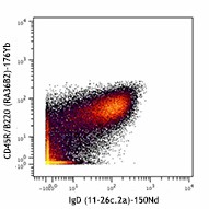
C57BL/6 mouse splenocytes stained with 176Yb-anti-CD45R/B220 (RA36B2) and 150Nd-anti-IgD (11-26c.2a). Lymphocytes are displayed in the analysis. Data provided by DVS Sciences.
| Cat # | Size | Price | Quantity Check Availability | ||
|---|---|---|---|---|---|
| 405737 | 100 µg | $94.00 | |||
Surface IgD is an important B cell differentiation marker.
Product Details
- Verified Reactivity
- Mouse
- Antibody Type
- Monoclonal
- Host Species
- Rat
- Formulation
- Phosphate-buffered solution, pH 7.2, containing 0.09% sodium azide and EDTA.
- Preparation
- The antibody was purified by affinity chromatography.
- Concentration
- 1.0 mg/ml
- Storage & Handling
- The antibody solution should be stored undiluted between 2°C and 8°C.
- Application
-
FC - Quality tested
CyTOF® - Verified - Recommended Usage
-
This product is suitable for use with the Maxpar® Metal Labeling Kits. For metal labeling using Maxpar® Ready antibodies, proceed directly to the step to Partially Reduce the Antibody by adding 100 µl of Maxpar® Ready antibody to 100 µl of 4 mM TCEP-R in a 50 kDa filter and continue with the protocol. Always refer to the latest version of Maxpar® User Guide when conjugating Maxpar® Ready antibodies.
- Application Notes
-
The 11-26c.2a antibody reacts with immunoglobulin D in all tested mouse haplotypes. The antibody binds membrane IgD expressed on most B cells. The 11-26c.2a antibody neither induces proliferation of splenic B cells nor induces B cell activation. Additional reported applications (for the relevant formats) include: immunohistochemical staining of acetone-fixed frozen sections2,3, and spatial biology (IBEX)10,11.
- Additional Product Notes
-
Maxpar® is a registered trademark of Standard BioTools Inc.
-
Application References
(PubMed link indicates BioLegend citation) -
- Nitschke L, et al. 1993. P. Natl. Acad. Sci. USA 90:1887. (FC)
- Weih D, et al. 2001. J. Immunol. 167:1909. (IHC)
- Koni PA, et al. 2001. J. Exp. Med. 193:741. (IHC)
- Ahuja A, et al. 2007. J. Immunol. 179:3351. (FC) PubMed
- Haynes NM, et al. 2007. J. Immunol. 179:5099. (FC)
- Good-Jacobson KL, et al. 2010. Nat. Immunol. 11:535. (FC) PubMed
- Tomayko MM, et al. 2010. J. Immunol. 185:7146. PubMed
- Park SY, et al. 2013. J. Immunol. 190:1094. PubMed
- Rouaud P, et al. 2014. J Exp Med. 211:975. PubMed
- Radtke AJ, et al. 2020. Proc Natl Acad Sci U S A. 117:33455-65. (SB) PubMed
- Radtke AJ, et al. 2022. Nat Protoc. 17:378-401. (SB) PubMed
- Product Citations
-
- RRID
-
AB_2563774 (BioLegend Cat. No. 405737)
Antigen Details
- Structure
- Ig family
- Distribution
-
B cells
- Function
- B cell differentiation
- Cell Type
- B cells
- Biology Area
- Immunology
- Gene ID
- 380797 View all products for this Gene ID
- UniProt
- View information about IgD on UniProt.org
Other Formats
View All IgD Reagents Request Custom ConjugationCompare Data Across All Formats
This data display is provided for general comparisons between formats.
Your actual data may vary due to variations in samples, target cells, instruments and their settings, staining conditions, and other factors.
If you need assistance with selecting the best format contact our expert technical support team.
-
FITC anti-mouse IgD
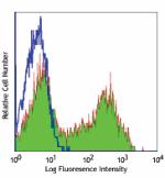
C57BL/6 mouse splenocytes stained with 11-26c.2a FITC -
PE anti-mouse IgD
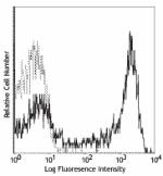
C57BL/6 splenocytes stained with 11-26c.2a PE -
Purified anti-mouse IgD
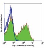
C57BL/6 mouse splenocytes stained with purified 11-26c.2a, f... -
PerCP anti-mouse IgD
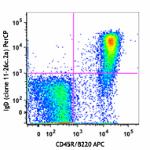
C57BL/6 mouse splenocytes were stained with CD45R/B220 APC a... 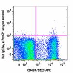
-
Biotin anti-mouse IgD
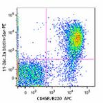
C57BL/6 splenocytes were stained with CD45R/B220 APC and bio... 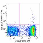
-
Brilliant Violet 711™ anti-mouse IgD
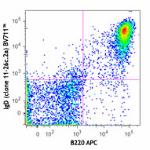
C57BL/6 mouse splenocytes were stained with B220 APC and IgD... 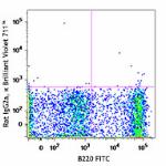
-
Alexa Fluor® 700 anti-mouse IgD
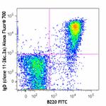
C57BL/6 mouse splenocytes were stained with B220 FITC and Ig... 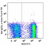
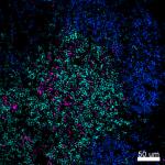
Confocal image of C57BL/6 mouse spleen sample acquired using... 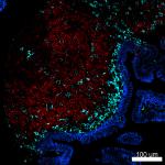
Confocal image of C57BL/6 mouse small intestine sample acqui... -
Alexa Fluor® 647 anti-mouse IgD
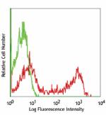
C57BL/6 splenocytes stained with 11-26c.2a Alexa Fluor® 647 
Paraformaldehyde-fixed (4%), 500 μm-thick mouse spleen secti... -
PerCP/Cyanine5.5 anti-mouse IgD
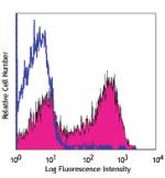
C57BL/6 splenocytes stained with 11-26c.2a PerCP/Cyanine5.5 -
Pacific Blue™ anti-mouse IgD
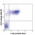
C57BL/6 splenocytes stained with 11-26c.2a Pacific Blue&trad... 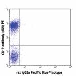
C57BL/6 splenocytes stained with rat IgG2a Pacific Blue&trad... -
APC anti-mouse IgD
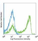
C57BL/6 splenocytes stained with 11-26c.2a APC -
APC/Cyanine7 anti-mouse IgD
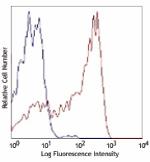
C57BL/6 splenocytes stained with 11-26c.2a APC/Cyanine7 -
Alexa Fluor® 488 anti-mouse IgD
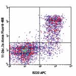
C57BL/6 splenocytes were stained with B220 APC and IgD (11-2... 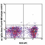
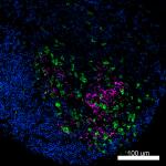
Mice were injected subcutaneously with sheep red blood cells... -
PE/Cyanine7 anti-mouse IgD

C57BL/6 splenocytes were stained with CD45R/B220 FITC and Ig... -
Brilliant Violet 650™ anti-mouse IgD
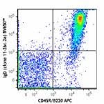
C57BL/6 splenocytes were stained with CD45R/B220 APC and IgD... 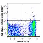
-
Brilliant Violet 510™ anti-mouse IgD
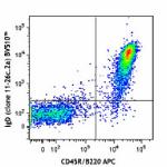
C57BL/6 splenocytes were stained with CD45R/B220 APC and IgD... 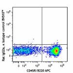
-
Brilliant Violet 421™ anti-mouse IgD
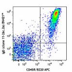
C57BL/6 splenocytes were stained with CD45R/B220 APC and IgD... 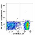
-
Brilliant Violet 605™ anti-mouse IgD
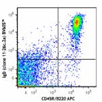
C57BL/6 splenocytes were stained with CD45R/B220 APC and IgD... 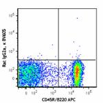
-
Purified anti-mouse IgD (Maxpar® Ready)
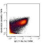
C57BL/6 mouse splenocytes stained with 176Yb-anti-CD45R/B220... -
Alexa Fluor® 594 anti-mouse IgD
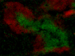
C57BL/6 mouse frozen spleen section was fixed with 4% parafo... 
Paraformaldehyde-fixed (4%), 500 μm-thick mouse spleen secti... -
PE/Dazzle™ 594 anti-mouse IgD
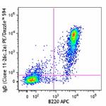
C57BL/6 mouse splenocytes were stained with B220 APC and IgD... 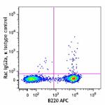
-
APC/Fire™ 750 anti-mouse IgD
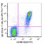
C57BL/6 mouse splenocytes were stained with CD45R/B220 FITC ... 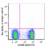
-
TotalSeq™-A0571 anti-mouse IgD
-
TotalSeq™-C0571 anti-mouse IgD
-
Spark NIR™ 685 anti-mouse IgD

C57BL/6 splenocytes were stained with anti-mouse CD45R/B220 ... -
TotalSeq™-B0571 anti-mouse IgD Antibody
-
Spark Violet™ 423 anti-mouse IgD

C57BL/6 splenocytes were stained with anti-mouse/human CD45R... -
PE/Cyanine5 anti-mouse IgD

C57BL/6 mouse splenocytes were stained with anti-mouse B220 ... -
Brilliant Violet 785™ anti-mouse IgD

C57BL/6 mouse splenocytes were stained with anti-mouse B220 ... -
Spark PLUS UV395™ anti-mouse IgD

C57BL/6 mouse splenocytes were stained with anti-mouse CD45R...
