IMPORTANT NOTICE: Our email addresses will soon change. Learn more >>

While some assays utilize antibodies to study cell health, proliferation, cell cycle or apoptosis, other types of experiments can rely on non-antibody based methods of assessment, often called non-antibody chemical probes. These are reagents that localize to an organelle or indicate health based on chemical characteristics like hydrophobicity, charge, size, and enzymatic activation.


Product Highlights


Image showing a cell, the organelles it contains within, and the reagents BioLegend provides to investigate them.

We have divided our chemical probes into tables based on their function or localization to help you choose the right reagent for your labeling application. Cell type suitability indicates whether this reagent labels live cells exclusively or whether it can also label the dead cells in a live cell culture or cells that have been previously fixed with buffers like paraformaldehyde or methanol. Sample suitability indicates whether this reagent can be used to label already fixed tissue sections or single cells in suspension or cell culture. Application indicates their suitability for being analyzed by flow cytometry, microscopy platforms or both. Fixation simply indicates whether the reagent, once used to label a cell sample, will be retained with a paraformaldehyde fixation.


Expand tables below to view chemical probes by localization and/or function.


  Spectra Functionality Cell Type Suitability Sample Suitability Application Fixation
Chemical Probe     Live Dead/ Fixed Tissue Cell Culture/ Single Cells Flow Cytometry Microscopy Retention with PFA Treatment
Apotracker™ Green View Spectra Apoptosis indicator    
Apotracker™ Tetra Alexa Fluor® 647 View Spectra Apoptosis indicator    
Annexin V Several formats available Apoptosis indicator      
7-AAD View Spectra Dead cell indicator    
Zombie UV387™ View Spectra Fixable cell death indicator    
Zombie UV™ View Spectra Fixable cell death indicator    
Zombie Violet™ View Spectra Fixable cell death indicator    
Zombie Aqua™ View Spectra Fixable cell death indicator    
Zombie Yellow™ View Spectra Fixable cell death indicator    
Zombie Green™ View Spectra Fixable cell death indicator    
Zombie B550™ View Spectra Fixable cell death indicator    
Zombie YG581™ View Spectra Fixable cell death indicator    
Zombie Red™ View Spectra Fixable cell death indicator    
Zombie R685™ View Spectra Fixable cell death indicator    
Zombie R718™ View Spectra Fixable cell death indicator    
Zombie NIR™ View Spectra Fixable cell death indicator    
Helix NP™ Blue View Spectra Viability indicator    
Helix NP™ Green View Spectra Viability indicator    
Helix NP™ NIR View Spectra Viability indicator    


  Spectra Functionality Cell Type Suitability Sample Suitability Application Fixation
Chemical Probe     Live Dead/ Fixed Tissue Cell Culture/ Single Cells Flow Cytometry Microscopy Retention with PFA Treatment
Flash Phalloidin™ Green 488 View Spectra F-actin staining     N/A
Flash Phalloidin™ Red 594 View Spectra F-actin staining     N/A


(+)    Cytoplasm


  Spectra Functionality Cell Type Suitability Sample Suitability Application Fixation
Chemical Probe     Live Dead/ Fixed Tissue Cell Culture/ Single Cells Flow Cytometry Microscopy Retention with PFA Treatment
Calcein-AM View Spectra Vitality indicator      
Calcein Red-AM View Spectra Vitality indicator      
Calcein Violet-AM View Spectra Vitality indicator      
Cellular Senescence Kit Red Exmax/Emmax 544, 567 nm Cellular senescence/cell cycle    
CFSE View Spectra Proliferation, cell tracking  
Tag-it Violet™ View Spectra Proliferation, cell tracking  


  Spectra Functionality Cell Type Suitability Sample Suitability Application Fixation
Chemical Probe     Live Dead/ Fixed Tissue Cell Culture/ Single Cells Flow Cytometry Microscopy Retention with PFA Treatment
Endoplasmic Reticulum Probe Green View Spectra Organelle staining      
Endoplasmic Reticulum Probe Red View Spectra Organelle staining      


(+)    Golgi Appartus


  Spectra Functionality Cell Type Suitability Sample Suitability Application Fixation
Chemical Probe     Live Dead/ Fixed Tissue Cell Culture/ Single Cells Flow Cytometry Microscopy Retention with PFA Treatment
Golgi Detection Probe Red Exmax/Emmax 
544, 570 nm
Organelle staining        


(+)    Lysosome


  Spectra Functionality Cell Type Suitability Sample Suitability Application Fixation
Chemical Probe     Live Dead/ Fixed Tissue Cell Culture/ Single Cells Flow Cytometry Microscopy Retention with PFA Treatment
Lysosome Probe Green View Spectra Organelle staining      
Lysosome Probe Orange View Spectra Organelle staining      
Lysosome Probe Red View Spectra Organelle staining      
Lysosome Probe Deep Red View Spectra Organelle staining      
Lysosome Probe NIR View Spectra Organelle staining      


(+)    Mitochondria


  Spectra Functionality Cell Type Suitability Sample Suitability Application Fixation
Chemical Probe     Live Dead/ Fixed Tissue Cell Culture/ Single Cells Flow Cytometry Microscopy Retention with PFA Treatment
JC-10 Mitochondrial Membrane Potential Kit View Spectra Depolarization/apoptosis indicator      
MitoSpy™ Green FM View Spectra Vitality indicator      
MitoSpy™ Orange CMTMRos View Spectra Vitality indicator    
MitoSpy™ Red CMXRos View Spectra Vitality indicator    
MitoSpy™ NIR DiIC1(5) View Spectra Vitality indicator      


(+)    Nucleus


  Spectra Functionality Cell Type Suitability Sample Suitability Application Fixation
Chemical Probe     Live Dead/ Fixed Tissue Cell Culture/ Single Cells Flow Cytometry Microscopy Retention with PFA Treatment
Helix NP™ Blue View Spectra Viability indicator    
Helix NP™ Green View Spectra Viability indicator    
Helix NP™ NIR View Spectra Viability indicator    
CytoPhase™ Violet View Spectra Cell cycle/viability indicator  
DRAQ5™ View Spectra Cell cycle/viability indicator  
DRAQ7™ View Spectra Cell cycle/viability indicator    
Propidium Iodide View Spectra Cell cycle/viability indicator    
DAPI View Spectra Nuclear stain/cell cycle    


  Spectra Functionality Cell Type Suitability Sample Suitability Application Fixation
Chemical Probe     Live Dead/ Fixed Tissue Cell Culture/ Single Cells Flow Cytometry Microscopy Retention with PFA Treatment
Autophagy Detection Probe Blue Exmax/Emmax 
330, 520 nm
Autophagy indicator        
Autophagy Detection Probe Green Exmax/Emmax 
485, 530 nm
Autophagy indicator        
Phagocytosis Detection Kit Red Exmax/Emmax 
570, 600 nm
Phagocytosis activity      


Typically tested by flow cytometry, proliferation can be assessed with probes that enter cells passively (such as CFSE-DA and Tag-it™ Violet). Inside the cell, esterase activity cleaves acetoxymethyl ester (AM) or diacetate (DA) groups from the probe, resulting in a charged fluorescent molecule that is retained by the intact membrane of the cell (left). Fluorescence intensity indicates the amount of esterase activity, which is dependent on the cell and its health status. Apoptotic and necrotic cells will exhibit lower esterase activity than healthy cells and dead cells will have even less, since only a small residual amount of leftover esterase may have been retained. As a cell divides, the resultant single cells will have half the fluorescent intensity of its parent cell (right).


How long the signal persists depends on the length of the experiment or how fast the cells are dividing. Higher concentrations of these probes can be toxic as they interfere with protein function. As such, titration of the dye is very important. If cell health is a concern in long-term cell tracking, consider using Zombie dyes, which only label cell surface primary amines of healthy cells and have little impact on cell health. However, Zombie dyes are not suitable for proliferation assays.  CFDA-SE, Tag-it Violet and the Zombie dyes are all retained in cells they have labeled upon fixation with paraformaldehyde to enable downstream antibody labeling for flow cytometry and imaging applications.

Tag-it Violet™


Mouse splenocytes stained with 5 µM Tag-it Violet™ without stimulation (left) or subsequently stimulated with 5 µg/mL ConA and 20 µg/mL IL-2 for 4 days (right).


Mouse spleen 72 hours after adoptive transfer of Tag-it Violet™-labeled splenocytes (purple). Nucleated cells are stained using 25 µM DRAQ™ (red). 

BrdU

BrdU (bromodeoxyuridine) assays are also a method of labeling proliferating cells. In culture or in vivo, BrdU is a nucleotide analog that can be fed to cells in culture media or via direct injection into an animal to be incorporated into the newly replicating DNA of dividing cells. The BrdU pulse time depends on the mitogen or the rate of cell division. This reveals which of the cells achieved division during the time of the BrdU pulse. While it will not give information about how many times a cell divided or the fate of all of the resultant daughter cells, it is a single cell flow cytometry application and thus additional parameters can be assessed on each cell like DNA content, phenotype of each cell or transcriptional factors and regulators that might have differentially responded to the mitogen.  As this application requires an anti-BrdU antibody, samples must be fixed and permeabilized for the antibody to gain access to the DNA for imaging.


BioLegend now offers Phase-Flow™ BrdU Kits designed for flow cytometric analysis and conveniently contain BrdU pulsing solution, an Anti-BrdU antibody, buffers, DNAse, PBS (with Ca2+/Mg2+), and dyes for total DNA staining (7-AAD and DAPI). 

Phase Flow™ Alexa Fluor® 647 anti-BrdU Antibody


Ramos cells loaded with BrdU for 1.5 hours (left) or with no load controls (right) stained using the Phase-Flow™ FITC BrdU Kit and DAPI.

Ki-67

Commonly looked at during proliferative stages of the cell cycle, Ki-67 is a nuclear protein involved in ribosomal RNA transcription. Expression occurs diffusely in the nucleus during the active phases of the cell cycle, G1, S and G2 phase, whereas in M phase or mitosis, the protein becomes localized to the surface of chromosomes. Ki-67 is not detectable in G0 or resting phase.


Con A-stimulated (3 days) BALB/c mouse splenocytes were fixed and permeabilized with 70% ethanol, then stained with Ki-67 (clone 16A8) Alexa Fluor® 647 (filled histogram) or rat IgG2a, κ Alexa Fluor® 647 isotype control (open histogram).


C57BL/6 mouse frozen intestine section was fixed, permeabilized, and blocked. Then the section was stained with anti-Ki-67 (clone 11F6) Alexa Fluor® 647 (red) and anti-E-cadherin (clone DECMA-1) Alexa Fluor® 594 (green) overnight at 4°C. Nuclei were counterstained with DAPI (blue). The image was captured with a 20X objective.

For time point assays, a microplate assay can be performed to give a broader view of the proliferation of cultured cells. Resazurin (Deep Blue Cell Viability™) and tetrazolium salts (LDH-Cytox™ Assay Kit) are probes that are used to determine the metabolic state of cells. The relative fluorescence or absorbance units of the microplate reader are plotted on a standard curve against number of live cells to give an overall view of rate of proliferation or cytotoxicity. Ideally, they should be paired with an impermeant nucleic acid stain to indicate the number of dead cells. Permeant nucleic acid stains can render a total cell count.

Deep Blue Cell Viability™


Detection of Baf3/CCR3 cells viability using Resazurin fluorescence measurement. The increasing cell numbers correlate with the increasing fluorescence.

The Deep Blue Cell Viability™ Kit is formulated to study cell proliferation and quantification. The extent of resazurin reduction and resorufin production is proportional to the number of metabolically active cells (live cells) present in the culture. The Deep Blue Cell Viability™ Kit can facilitate cytotoxicity studies in high-throughput screening, as well as the evaluation of new drugs and chemicals.

LDH-Cytox Assay™ Kit

LDH-Cytox Assay™ Kit can be used for determination of cytotoxicity by measuring lactate dehydrogenase activity released from damaged cells. LDH catalyzes dehydrogenation of lactate to pyruvate thereby reducing NAD+ to NADH. NADH reduces water-soluble tetrazolium salt in the presence of an electron mediator to produce an orange formazan dye.


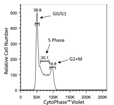
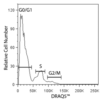
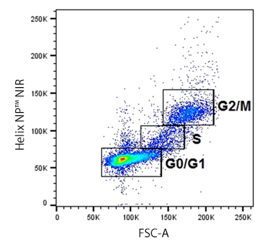
Cellular division for any cell type is dependent on the inherent function, location and the response of cells to repair, apoptosis, or death. To divide, cells must duplicate a copy of their DNA, increase mitochondrial density, and assemble/synthesize microtubules during interphase. G2 is a checkpoint stage of interphase where the cell has two sets of dsDNA and must commit to mitosis. Mitosis is the actual division stage where two daughter cells are created. There can be symmetrical or asymmetrical division depending on the cell and tissue type. Mitosis can be further subdivided into prophase, metaphase, anaphase, and telophase as the nucleus undergoes division and chromatids are pulled away from one another. After mitosis, the cell will undergo the G0/G1 phase when the cells rest to become ready for the next round of replication.

cycle

During G0/G1, S, and G2 phases, some fluorogenic nucleic acid stains selectively bind dsDNA stoichiometrically to give us measurements of DNA mass based on fluorescence intensity. Cell-permeant nucleic acid stains can be used in instances where cells will be analyzed live. Propidium IodideDAPIDRAQ5™DRAQ7™CytoPhase™ VioletHelix NP™ NIR,  Helix NP™ Blue, and Helix NP™ Green can all be used to stain fixed cells for cell cycle analysis. However, only CytoPhase™ Violet and DRAQ5™ can be used to assess DNA content of live cells.


For additional reagents (including antibodies) on cell cycle analysis or DNA replication, check here.

Live and Dead Cell Cycle Analysis

CytoPhase™ Violet


Live Ramos cells treated with 5 µM CytoPhase™ Violet dye for 90 minutes at 37°C. 

DRAQ5™


Live C57BL/6 mouse bone marrow cells were stained with DRAQ5™.
 

Helix NP™ NIR


C57BL/6 mouse thymus cells were fixed using 70% chilled ethanol. The cells were incubated for one hour at -20°C, washed, then stained with Helix NP™ NIR at 5 µM.

Impermeant Nucleic Acid Stains

Impermeant nucleic acid stains only gain entrance to the cell if membrane integrity is compromised. Dead cells will stain brightly and live cells will not stain at all. Apoptotic cells, however, can also allow entrance of certain otherwise impermeant nucleic acid stain at late stages, like DAPI and Helix NP™. Assessing multiple markers for different characteristics of apoptosis are important. For example, the most commonly used marker of apoptosis, Annexin V, will stain phosphatidylserine (PS) residues that have been translocated to the cell surface during the early to mid stages of apoptosis. But, it will also stain the intracellular-facing PS due to the loss of membrane integrity in death. Therefore, an impermeant nucleic acid stain must be employed to distinguish cells dying from apoptosis from those dead from necrosis. Since some nucleic acid stains can start to stain late stage apoptotic cells, the best impermeant nucleic acid stains for dead cells are Propidium IodideHelix NP™ BlueHelix NP™ Green, and Helix NP™ NIR.

Helix NP™ NIR

Helix NP™ NIR

One day old C57 mouse splenocytes were stained with Helix NP™ NIR (filled histogram). Cells alone, without Helix NP™ NIR staining, are also shown (open histogram).

Helix NP™ Green

Helix NP™ Green

One day old C57BL/6 mouse splenocytes were stained with 5 µM Helix NP™ Green and APC Annexin V with no wash.

Zombies Dyes

Zombie dyes, which are fully explained on our live/dead webpage, exploit the permeability of the cell membrane to indicate live versus dead, but do not bind to DNA. Zombie dyes are a family of amine-reactive dyes that do not passively cross the cell membrane due to their valency. Only if the cell is compromised can these small molecules passively cross. Inside the cell, they covalently conjugate an abundance of intracellular proteins and the signal is retained with paraformaldehyde fixation. Titration of this process is critical since the goal is to use the lowest possible concentration of the Zombie dye to achieve a bright intracellular staining (dead cell status) with low residual cell surface staining (live cell status/background staining). Zombie dyes are most commonly used in flow cytometry, but when optimized, can also be applied to imaging cell in culture. Other metabolic probes can also exploit the permeability of the cell membrane to assess live/dead status and are described in the section for vitality indicators.


(Left) Impermeant nucleic acid stains can only stain DNA when the membrane is compromised. (Right) Zombie dyes bind to primary amines on proteins. Live cells receive low level surface staining, while dead cells stain at a much brighter fluorescence level due to the exposure of internal proteins.

Zombies in Microscopy

Zombie Violet™

Zombie Green™

Zombie Red™

zombie image

HeLa cells were treated with 20% EtOH for 20 seconds, washed twice with PBS, and then were left to recover for five minutes with cell culture media in 37°C. The cells were stained with Zombie Violet™ (magenta, left), Zombie Green™ (green, middle), or Zombie Red™ (red, right) at a 1:1000 dilution for 15 minutes and then fixed with 1% PFA for ten min. Nuclei were counterstained with DRAQ5™ (blue) for five min. The image was captured with 40X objective.

Zombies in Flow Cytometry

Zombie UV™ Zombie Violet™ Zombie Aqua™ Zombie Yellow™ Zombie Green™ Zombie Red™ Zombie NIR™
zombie image 2

One day old C57BL/6 mouse splenocytes were stained with Zombie dyes as indicated and analyzed before fixation (blue/purple) or after fixation and permeabilization (red). Cells alone, without Zombie staining, are indicated in black.

Reagent Equivalent Channel Optimal Excitation Laser Excitation/Emission Max
Zombie UV™ DAPI UV 355 nm 362 nm/459 nm
Zombie Violet™ BV421™, Pacific Blue Violet 405 nm 400 nm/423 nm
Zombie Aqua™ BV510™ Violet 405 nm 382 nm/510 nm
Zombie Yellow™ BV570™ Violet 405 nm 396 nm/572 nm
Zombie Green™ FITC Blue 488 nm 491 nm/515 nm
Zombie B550™ N/A Blue 488 nm 516 nm/540 nm
Zombie YG581™ N/A Yellow/Green 532/561 nm 565 nm/581 nm
Zombie Red™ PE/Texas Red® Blue 488 nm or Yellow/Green 532/561 nm 600 nm/624 nm
Zombie R685™ N/A Red 633 nm 660 nm/685 nm
Zombie R718™ N/A Red 633 nm 697 nm/711 nm
Zombie NIR™ APC/Cyanine7 Red 633 nm 719 nm/746 nm

Note that for some Zombie Dyes, a direct equivalent channel is not listed due to the nature of spectral unmixing cytometers.

Apotracker™

Apotracker™ is a family of fluorogenic probes that detects externalized phosphatidylserine (PS) residues on apoptotic cells in a calcium-independent manner. Traditional apoptosis detection probes, such as Annexin V, require the presence of calcium, which can affect cell viability and marker expression. Apotracker exhibits a linear relationship with Annexin V staining and does not require a specialized buffer. It can also be useful for microscopy assays where it presents less background staining than traditional probes.

Annexin V

Apoptosis is a death process defined by the internal degradation of cellular components without the instigation of a systemic inflammation response. Annexin V is one of the most common probes used to examine early-to-mid stages of apoptosis. Annexin V is a protein that recognizes phosphatidylserine (PS), which is commonly found on the cytosolic side of the membrane in healthy cells. However, in dying or apoptotic cells, PS translocates to the extracellular side of the membrane, exposing it and making it available for binding by Annexin V in a calcium-dependent manner. BioLegend provides several fluorophore-conjugated Annexin V options, as well as an array of impermeant nucleic acid stains to be used for distinguishing apoptotic from necrotic cells, and the Annexin V Binding Buffer required for the calcium-dependent binding of the annexin to PS.

LEAF™ Purified Anti-human CD95 (Fas) Antibody


Human T-cell leukemia cell line, Jurkat, treated (top) or non-treated (bottom) with BioLegend's LEAF™ purified anti-human CD95 (clone EOS9.1) mAb (Cat. No. 305704) for 4 hours, then stained with FITC Annexin V Apoptosis Detection Kit with 7-AAD (Cat. No. 640922).


Activated Caspases

Caspases are an endoprotease family that work as an orchestra of signaling molecules involved in inflammation and apoptosis by cleaving and degrading intracellular components to control the disposal of cellular debris. Dysregulation of caspases underlies diseases like cancer, developmental abnormalities, and autoimmune inflammatory disorders. Caspases integral to the “death cascade” are activated in a middle stage of apoptosis when they can form dimers and be cleaved into large and small subunits of differing function. For example, when a cell begins to enter apoptosis, it releases cytochrome c, a protein involved in mitochondrial respiration, from the intermembrane space. Cytochrome c complexes with Apaf-1 to form an apoptosome that recruits and activates procapase-9, leading to the further activation of caspase-3.

caspase

Explore the full interactive poster here

Activated caspase-3 is involved in the signaling cascade responsible for apoptosis execution. Caspase-3 has many downstream targets to cleave including PARP (poly ADP-ribose polymerase). PARP is involved in repairing nicked DNA and keeping cells viable. Cleavage of PARP inactivates this function and facilitates apoptosis and cell death. This cascade of cytochrome c, caspase-9, caspase-3 and PARP are some of the more popular markers to follow when assessing the progression of apoptosis. A great review of the function of individual signaling molecules involved in apoptosis can be found here.

Initiator Caspases
Caspase 2, 8, 9, 10
Executioner Caspases
Caspase 3, 6, 7
Inflammation Caspases
Caspase 1,4,5,12,14

Bcl-2 and Bax

The Bcl-2 family of proteins inhibit apoptosis by interfering with the function of pro-apoptotic proteins at the mitochondrial outer membrane, thus promoting cell survival. Overexpression of anti-apoptotic proteins can result in a lack of the necessary regulated cell death needed to maintain homeostasis, promoting the development of cancer and a lack of necessary immuno-surveillance in autoimmune disorders. A Bcl-2 homologous protein called Bax promotes cell death by competing with Bcl-2. While Bax-Bax homodimers act as potent apoptosis inducers. Antibodies against the Bcl-2 and Bax families are useful when correlated with other markers of cell health and apoptosis and when it is possible to detect relative differences in abundance of the expression of these markers.


There are, of course, several other markers that can be analyzed to study apoptosis, tumor suppressors, or cell death. You can find more reagents here.

Esterase-Dependent Probes

Other functional probes can also assess live/dead status depending on their cellular vitality. Long-term cell tracking probes like CFSE help to indicate a cell’s health as it relies on its esterase activity. The simplest example of cell vitality, however, is Calcein-AM, Calcein Red-AM and Calcein Violet-AM. These are simple fluorogenic esterase probes that are passively cell-permeant and then subsequently retained by the cell when the probe becomes charged upon conversion. The brighter the signal, the more abundant the esterase activity and the healthier the cell. As cells die, whether by necrosis or apoptosis, the esterase activity will become reduced until no activity remains. These probes can be used to track the cells for as long as the signal persists. However, they are considered short-term trackers since they do not bind to internal proteins like CFSE does, and most cells will eventually start to pump out these probes naturally, or actively in the case of cells that are multidrug resistant (MDR).

Calcein-AM


Fresh (left) or day-old C57BL/6 splenocytes (right) were stained with 0.01 µM Calcein AM and a cell-impermeant nucleic acid dye, SYTOX™ Red (colored). Black figure represents unstained splenocytes.

Mitochondrial Respiration

Similarly, mitochondrial respiration and thus the polarization of the mitochondrial membrane are also indicators of cell health. Probes like MitoSpy™ Orange CMTMRos and MitoSpy™ Red CMXRos are both live cell-permeant fluorogenic probes that localize to the mitochondrial membrane based on its strong polarization in healthy, respiring cells. The most common application of MitoSpy™ probes is simple mitochondrial localization in live cells for imaging applications. However, when used in conjunction with other probes for cell health, like Annexin V, Calcein-AM, or impermeant nucleic acid stains, the neutralization of potential across the mitochondrial membrane can be one of the first indications that a cell is entering apoptosis. Learn more with our MitoSpy™ webpage.

MitoSpy™ Orange


Human T-cell leukemia cell line, Jurkat, was treated for five hours with LEAF™ purified anti-CD95 (clone EOS9.1), then stained with an impermeant nucleic acid stain, Annexin V APC, and MitoSpy™ Orange CMTMRos. Data shown was gated on live cells.


HeLa cells were stained with 250 nM of MitoSpy™ Orange CMTMRos (yellow) for 20 minutes, fixed with 4% PFA for ten minutes, and permeabilized with 0.1% Triton X-100 for ten minutes. Then the cells were stained with anti-Cytochrome C Alexa Fluor® 647 (red) and counterstained with DAPI (blue). All three images were merged in the bottom right panel. The image was captured with a 60x objective.

MitoSpy™ Red


Human T-cell leukemia cell line, Jurkat, was treated for five hours with LEAF™ purified anti-CD95 (clone EOS9.1), then stained with an impermeant nucleic acid stain, Annexin V Alexa Fluor® 488, and MitoSpy™ Red CMXRos. Data shown was gated on live cells.


NIH3T3 cells were stained with 100 nM of MitoSpy™ Red CMXRos (red) for 20 minutes at 37°C, fixed with 1% PFA for ten minutes at room temperature, and permeabilized with 1X True Nuclear™ Perm Buffer for ten minutes at room temperature. Then the cells were stained with Flash Phalloidin™ NIR 647 (green) for 20 minutes at room temperature and counterstained with DAPI (blue). The image was captured with a 60x objective.


Login / Register
Remember me
Forgot your password? Reset password?
Create an Account
My Cart (0 items)