
 Login / Register


- Clone
- N152B/23 (See other available formats)
- Regulatory Status
- RUO
- Other Names
- Voltage-dependent anion-selective channel protein 1, porin 31HL, porin 31HM, plasmalemmal porin, outer mitochondrial membrane protein porin 1, PORIN, VDAC-1
- Previously
-
Covance Catalog# MMS-5205
- Isotype
- Mouse IgG2a, κ
- Ave. Rating
- Submit a Review
- Product Citations
- 8 publications


| Cat # | Size | Price | Quantity Check Availability | Save | ||
|---|---|---|---|---|---|---|
| 820702 | 25 µL | 83 CHF | ||||
| 820701 | 100 µL | 165 CHF | ||||
Voltage-dependent anion-selective channel protein 1 (VDAC1, also known as outer mitochondrial membrane protein porin 1, porin 31HL, and plasmalemmal porin) is a voltage-dependent anion channel and is the main calcium ion transport channel and plays a role in regulating the citric acid cycle and ROS generation. VDAC1 is implicated in cancer and neurodegenerative diseases through its ability to elevate ROS production.
Product DetailsProduct Details
- Verified Reactivity
- Human, Mouse, Rat
- Antibody Type
- Monoclonal
- Host Species
- Mouse
- Immunogen
- This monoclonal antibody was raised against a fusion protein corresponding to amino acids 1-283 (full-length) of human VDAC1.
- Formulation
- Phosphate-buffered solution.
- Preparation
- The antibody was purified by affinity chromatography.
- Concentration
- 1 mg/ml
- Storage & Handling
- This antibody should be handled aseptically as it is free of preservatives such as Sodium Azide. Store this antibody undiluted between 2°C and 8°C. Please note the storage condition for this antibody has been changed from -20°C to between 2°C and 8°C. You can also check the vial label or CoA to find the proper storage conditions.
- Application
-
WB - Quality tested
IHC-P - Verified - Recommended Usage
-
Each lot of this antibody is quality control tested by Western blotting. For Western blotting, the suggested use of this reagent is 1.0 - 5.0 µg per ml. For immunohistochemistry of formalin-fixed paraffin-embedded tissue, a concentration range of 1.0 - 5.0 µg/ml is suggested. It is recommended that the reagent be titrated for optimal performance for each application.
- Application Notes
-
This antibody is effective in immunoblotting (WB) and immunohistochemistry (IHC).
- Product Citations
-
- RRID
-
AB_2750531 (BioLegend Cat. No. 820702)
AB_2564843 (BioLegend Cat. No. 820701)
Antigen Details
- Structure
- VDAC1 is a 283 amino acid protein with a molecular weight of 30 kD.
- Biology Area
- Cell Biology, Mitochondrial Function, Neuroscience, Neuroscience Cell Markers
- Molecular Family
- Mitochondrial Markers
- Gene ID
- 7416 View all products for this Gene ID
- UniProt
- View information about VDAC1 on UniProt.org


Related FAQs
Other Formats
View All VDAC1 Reagents Request Custom Conjugation| Description | Clone | Applications |
|---|---|---|
| Purified anti-VDAC1 | N152B/23 | WB,IHC-P |
Customers Also Purchased
Compare Data Across All Formats
This data display is provided for general comparisons between formats.
Your actual data may vary due to variations in samples, target cells, instruments and their settings, staining conditions, and other factors.
If you need assistance with selecting the best format contact our expert technical support team.
-
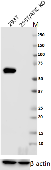
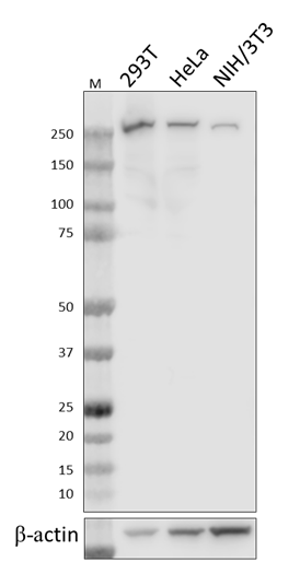
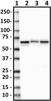
Purified anti-VDAC1

Western blot of anti-VDAC1 antibody (clone N152B/23). Lane 1... 
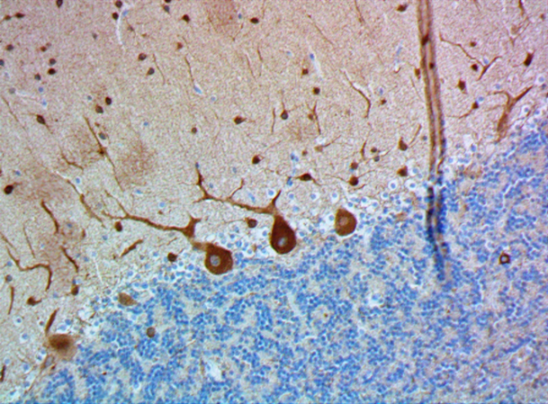
IHC staining of anti-VDAC1 antibody (clone N152B/23) on form... 
IHC staining of anti-VDAC1 antibody (clone N152B/23) on form... 
IHC staining of anti-VDAC1 antibody (clone N152B/23) on form... 
IHC staining of anti-VDAC1 antibody (clone N152B/23) on form...


Follow Us